1- Definition and Classification
2-Pathophysiology
3-Triggers and Causes
4- Clinical Features and Timeline
5– Serum Sickness–Like Reactions (SSLR)
6- Schistosomiasis and ‘Serum Sickness’
7- Laboratory Findings
8- Laboratory Findings
9- Differential Diagnosis
10- Treatment and Prognosis
11- Prevention Strategies
Serum sickness is a type III hypersensitivity reaction caused by immune complexes formed after exposure to foreign proteins.
It is a systemic immune reaction, distinct from immediate allergic (type I) reactions.

Antigen–antibody complexes form in circulation.
These complexes deposit in blood vessels, joints, kidneys.
Complement activation → vasculitis, inflammation, and tissue damage.
Common causes include:
Heterologous antiserum (e.g. equine anti-venom, anti-diphtheria)
Monoclonal antibodies (e.g. rituximab, infliximab)
Penicillin (rare but classic)
Infections (e.g. schistosomiasis, particularly in acute stages)
Typical onset: 7–12 days after antigen exposure.
Symptoms:
Fever
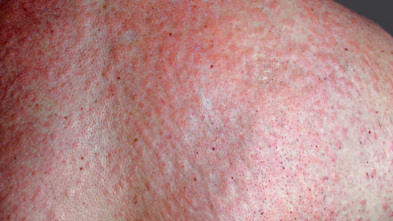
Urticarial or morbilliform rash
Arthralgia and arthritis
Lymphadenopathy
Myalgia, malaise
Occasionally: nephritis, neurological symptoms

Similar symptoms but not true type III hypersensitivity.
Often seen in children after antibiotics (e.g. cefaclor).
Less complement consumption, no circulating immune complexes.
Acute phase (Katayama fever) of S. mansoni / japonicum can mimic serum sickness:
Fever, rash, myalgia, eosinophilia.
Due to immune complex formation and systemic inflammation
↑ ESR and CRP
↓ Complement levels (C3, C4)
Mild proteinuria or haematuria (if renal involvement)
Circulating immune complexes (if tested)
History of exposure to foreign protein or biologic agent
Onset of typical symptoms in 1–2 weeks
Supportive labs (↑ inflammatory markers, ↓ complement)
Anaphylaxis (immediate, type I, IgE-mediated)
Drug rash (exanthema)
Viral exanthems
Systemic autoimmune diseases (e.g. SLE)
Withdraw the offending agent
Supportive care:
Antihistamines for rash
NSAIDs or corticosteroids for severe symptoms
Usually self-limiting (resolves in 1–2 weeks)
Monitor renal function if glomerulonephritis suspected
Avoid re-exposure to known triggers
Caution with repeat use of non-human antiserum
Screen for hypersensitivity before administering biologic drugs