Lymphadenopathy
Content of This Page
1- Introduction
2- General Classification & Causes
3- Pathophysiology
4- Clinical Features
5- Investigations
6- Treatment
7- Prognosis & Follow-up
8- Disease-Specific Presentations
Introduction
What is Lymphadenopathy?
Abnormal enlargement or alteration of lymph nodes due to infection, inflammation, or malignancy.
Types
Localised – one region (e.g. cervical)
Generalised – multiple non-contiguous areas
Common Causes
Infectious – viral (EBV, HIV), bacterial (TB), parasitic
Neoplastic – lymphoma, leukaemia, metastases
Autoimmune – SLE, rheumatoid arthritis
Drug-induced – phenytoin, allopurinol
Other – sarcoidosis, storage disorders
Clinical Importance
May be benign or a sign of serious disease
Key: assess size, consistency, tenderness, mobility
Watch for red flags: hard, fixed, painless nodes + systemic symptoms (fever, night sweats, weight loss)
General Classification & Causes
1. Classification
By Distribution
Localised lymphadenopathy
Involves a single region (e.g. cervical, axillary, inguinal)
Often due to local infection or malignancy
Generalised lymphadenopathy
Involves two or more non-contiguous areas
Suggests systemic disease (e.g. viral infections, autoimmune disease, malignancy)
2. General Causes of Lymphadenopathy
A. Infectious
Viral
Epstein–Barr virus (EBV) – infectious mononucleosis
Cytomegalovirus (CMV)
HIV – persistent generalised lymphadenopathy
Rubella, measles, adenovirus
Bacterial
Tuberculosis (matted, may form sinus tracts)
Secondary to bacterial infections of skin, throat, or soft tissues
Syphilis, cat scratch disease (Bartonella)
Parasitic/Fungal
Toxoplasmosis
Histoplasmosis
B. Neoplastic
Haematological malignancies
Hodgkin lymphoma (typically painless, rubbery nodes)
Non-Hodgkin lymphoma
Chronic lymphocytic leukaemia (CLL)
Metastatic cancers
Head and neck cancers → cervical nodes
Breast cancer → axillary nodes
Testicular or pelvic cancers → inguinal or supraclavicular nodes
C. Autoimmune/Inflammatory
Systemic lupus erythematosus (SLE)
Rheumatoid arthritis
Sarcoidosis
Often bilateral hilar lymphadenopathy (BHL)
D. Drug-Induced
Phenytoin (most classic)
Allopurinol
Hydralazine
3. Clinical Note
Tender, soft, mobile nodes → typically reactive/infectious
Hard, fixed, non-tender nodes → suspicious for malignancy
Supraclavicular nodes are particularly concerning and should always be investigated
Pathophysiology
1. Reactive (Hyperplastic) Lymphadenopathy
Triggered by antigenic stimulation from infection or inflammation
Follicular hyperplasia: expansion of B-cell zones
Paracortical hyperplasia: T-cell activation
Sinus histiocytosis: seen in draining nodes from cancers or severe inflammation
Common in viral infections (e.g. EBV, HIV) and autoimmune diseases (e.g. SLE)
2. Infective Lymphadenopathy
Bacterial or granulomatous infections (e.g. TB, cat-scratch disease) cause:
Suppuration: pus formation within node (abscess)
Granuloma formation: chronic inflammation with macrophages, caseation (TB)
Nodes may become matted, tender, or fluctuant
3. Neoplastic Lymphadenopathy
a. Lymphoid Malignancies
Hodgkin and Non-Hodgkin lymphoma involve neoplastic proliferation of lymphocytes
Architecture is replaced by malignant cells
Results in painless, firm, rubbery, non-tender nodes
b. Metastatic Cancer
Tumour cells spread via lymphatics to regional nodes
Leads to firm, fixed nodes with loss of normal structure
Often found in supraclavicular (Virchow’s node), axillary, or inguinal areas
4. Infiltrative Causes
Storage diseases (e.g. Gaucher’s disease) or sarcoidosis
Non-malignant infiltration of immune cells or abnormal proteins
Results in generalised enlargement of nodes with preserved architecture
5. Drug-Induced Lymphadenopathy
Certain drugs (e.g. phenytoin) cause immune stimulation or pseudo-lymphoma reactions
Pathology shows lymphoid hyperplasia without malignancy
Clinical Features
1. General Presentation
Lymphadenopathy may present as:
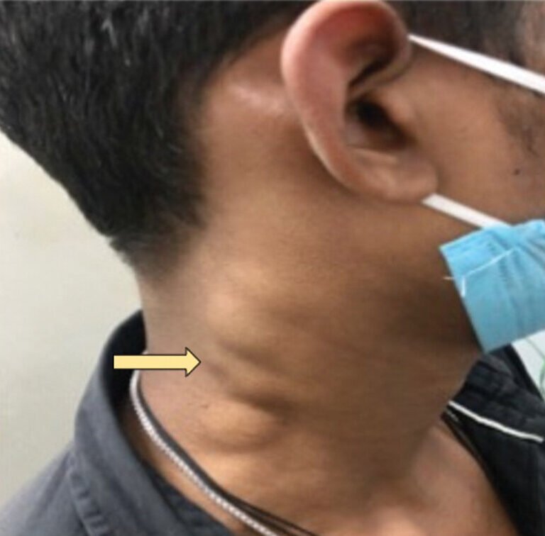
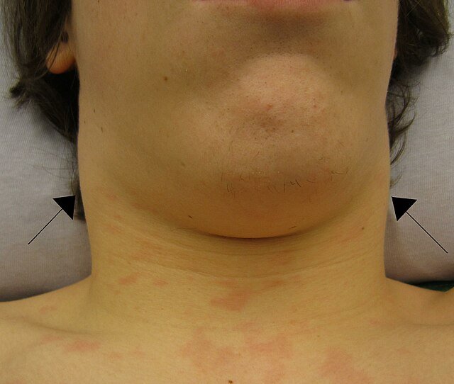
Localised swelling (e.g. neck, axilla, groin)
Generalised enlargement (2 or more regions involved)
The nodes themselves may be:
Palpable (size >1 cm often clinically significant)
Visible (if superficial and enlarged)
Tender or non-tender
2. Node Characteristics to Assess
| Feature | Suggestive of |
|---|---|
| Size | >1 cm significant (especially if persistent) |
| Tenderness | Tender → likely inflammatory (e.g. viral/bacterial) |
| Consistency | Firm/hard → malignancy; Soft → benign/reactive |
| Mobility | Fixed → infiltrative/malignant; Mobile → benign |
| Matted nodes | TB, lymphoma |
| Fluctuation | Abscess formation in bacterial infection |
3. Associated Symptoms
Systemic features (suggest serious pathology):
Fever
Night sweats
Weight loss
Fatigue
These are known as “B symptoms” in lymphoma.
Local features:
Redness, warmth, and pain (suggest acute infection)
Skin changes, draining sinuses (e.g. in tuberculous lymphadenitis)
Organ-specific clues:
Sore throat → cervical nodes (e.g. EBV, tonsillitis)
Genital ulcers or discharge → inguinal nodes
Pulmonary symptoms (cough, wheeze) → hilar or mediastinal nodes (e.g. TB, sarcoidosis)
4. Anatomical Site Clues
| Site | Common Causes |
|---|---|
| Cervical | Viral URTI, EBV, lymphoma, TB |
| Axillary | Breast cancer, cat-scratch disease, lymphoma |
| Inguinal | STIs, skin infections, lymphoma |
| Supraclavicular | Always suspicious for malignancy (e.g. gastric, lung) |
| Hilar/mediastinal | Sarcoidosis, lymphoma, TB |
Investigations
1. Clinical Goals of Investigation
Distinguish benign (reactive/infectious) vs. malignant (neoplastic/metastatic) causes
Identify systemic disease (e.g. infection, autoimmune disorder)
Determine need for biopsy or specialist referral
2. Initial Clinical Assessment
History: duration, associated symptoms (fever, weight loss, infections, drug use)
Examination: node size, site, tenderness, consistency, fixation
Check for organomegaly, skin lesions, ENT source, and site-specific clues
3. Basic Laboratory Tests
| Test | Purpose |
|---|---|
| Full blood count (FBC) | Anaemia, leukocytosis (infection), lymphocytosis (CLL), cytopenias (bone marrow infiltration) |
| ESR/CRP | Marker of inflammation or malignancy |
| Liver and renal function tests | Assess organ involvement |
| HIV serology | Rule out chronic viral infection |
| EBV/CMV serology | If infectious mononucleosis suspected |
| Tuberculin skin test or IGRA | Screen for TB |
| Autoimmune markers (ANA, RF) | In suspected SLE or RA |
4. Imaging
| Imaging | Indication |
|---|---|
| Chest X-ray | Mediastinal or hilar lymphadenopathy (e.g. TB, sarcoidosis, lymphoma) |
| Ultrasound | Assess nodal size, structure (especially in axilla, neck, groin) |
| CT/MRI | Define extent, especially for deep or intra-abdominal nodes |
| PET-CT | In staging lymphoma or identifying occult malignancy |
5. Definitive Diagnostic Tests
Fine Needle Aspiration Cytology (FNAC)
Quick, minimally invasive
May suggest malignancy or granulomatous inflammation
Not sufficient to diagnose lymphoma on its own
Excisional Biopsy
Gold standard if malignancy suspected
Preserves architecture → needed for lymphoma subtyping
Indicated if:
Node >2 cm and persists >4–6 weeks
Hard, fixed, non-tender
Supraclavicular location
Accompanied by B symptoms
6. Microbiological Testing
Culture of aspirate or biopsy material
Stain for acid-fast bacilli (if TB suspected)
PCR for specific pathogens (e.g. TB, toxoplasma)
Treatment
1. Reactive or Infectious Lymphadenopathy
a. Viral Infections (e.g. EBV, CMV, HIV)
Supportive care: rest, fluids, antipyretics
Avoid antibiotics unless secondary infection suspected
Monitor for resolution (usually within 2–6 weeks)
b. Bacterial Infections (e.g. Staphylococcus, Streptococcus)
Empirical antibiotics based on likely source (e.g. throat, skin)
Drain abscesses if fluctuant (especially in children)
c. Tuberculous Lymphadenitis
Standard anti-TB regimen (e.g. RIPE: Rifampicin, Isoniazid, Pyrazinamide, Ethambutol for 6 months)
Surgical drainage if suppuration or sinus formation
2. Malignant Lymphadenopathy
a. Lymphoma (Hodgkin or Non-Hodgkin)
Chemotherapy ± radiotherapy
E.g. ABVD for Hodgkin lymphoma
Biopsy is essential for histological diagnosis and staging
PET-CT for treatment monitoring
b. Metastatic Cancer
Treat primary malignancy (e.g. lung, breast, GI, head and neck)
Surgery, chemotherapy, or targeted therapy based on tumour type
3. Autoimmune Lymphadenopathy (e.g. SLE, RA)
Treat underlying disease with:
Immunosuppressants (e.g. corticosteroids, hydroxychloroquine, methotrexate)
Disease-modifying drugs based on rheumatological diagnosis
4. Drug-Induced Lymphadenopathy
Withdraw offending agent (e.g. phenytoin, allopurinol)
Monitor for resolution over weeks
Biopsy if persistence beyond 4–6 weeks or concerning features
5. Supportive Measures
Analgesia for tender nodes
Warm compresses may reduce local inflammation in reactive causes
Regular follow-up for unresolved or progressive nodes
Prognosis & Follow-up
1. Prognosis
Benign/reactive lymphadenopathy (e.g. viral infections)
Excellent prognosis
Resolves spontaneously within 2–6 weeks
No long-term consequences
Infective causes (bacterial, TB)
Good prognosis with prompt antibiotic or anti-TB therapy
Risk of sinus formation or recurrence in TB
Malignancy-associated lymphadenopathy
Prognosis depends on:
Histological type (e.g. Hodgkin vs. high-grade NHL)
Stage at diagnosis
Response to chemotherapy
Hodgkin lymphoma has a high cure rate, especially in early stages
Autoimmune causes
Flares may cause intermittent lymphadenopathy
Prognosis tied to disease control (e.g. SLE, RA)
2. Follow-up Principles
a. General Monitoring
Reassess node size, tenderness, and new symptoms
Most benign/reactive nodes resolve within 4–6 weeks
If persistent, consider imaging or biopsy
b. Red Flags Prompting Further Action
Node >2 cm and persisting beyond 6 weeks
Hard, fixed, or supraclavicular nodes
Associated systemic symptoms (fever, weight loss, night sweats)
c. Disease-Specific Follow-up
TB lymphadenitis: Monitor resolution on therapy; assess for drug resistance if relapse
Lymphoma: Serial PET-CT, blood counts, and physical exam as per protocol
Autoimmune disease: Track inflammatory markers, symptoms, and response to DMARDs
3. Patient Education
Reassure when reactive cause is likely
Educate on red flag symptoms
Encourage follow-up if nodes persist or worsen
Disease-Specific Presentations
1. Infectious Mononucleosis (EBV/CMV)
Generalised lymphadenopathy (esp. posterior cervical)
Nodes: soft, tender, mobile
Associated features: fever, sore throat, splenomegaly
Labs: atypical lymphocytosis, positive Monospot or EBV serology
2. HIV Infection
Persistent generalised lymphadenopathy (PGL): non-tender, symmetrical
Often affects cervical, axillary, and inguinal nodes
Nodes may persist for months
Consider opportunistic infections (e.g. TB, lymphoma, Kaposi’s sarcoma) in later stages
3. Tuberculous Lymphadenitis
Common in cervical or supraclavicular regions
Nodes: firm → matted → fluctuant → may form sinus tracts
Known as “scrofula” when in the neck
Systemic symptoms: fever, night sweats, weight loss
Diagnosis: aspirate or biopsy → caseating granulomas, AFB stain or TB PCR
4. Hodgkin Lymphoma
Painless, rubbery, firm nodes (often cervical or mediastinal)
May have B symptoms: fever, night sweats, weight loss
Alcohol-induced node pain is a classic (but rare) clue
Excisional biopsy shows Reed–Sternberg cells
5. Non-Hodgkin Lymphoma (NHL)
Can be localised or generalised
May involve extranodal sites (e.g. GI tract, CNS)
Nodes: firm, non-tender, fixed
Often more rapid progression and systemic symptoms than Hodgkin
6. Sarcoidosis
Bilateral hilar lymphadenopathy (BHL) ± peripheral nodes
Non-caseating granulomas on biopsy
Associated features: uveitis, erythema nodosum, arthritis, fatigue
May present as part of Löfgren’s syndrome (BHL + erythema nodosum + arthritis)
7. Systemic Lupus Erythematosus (SLE)
Mild, symmetrical lymphadenopathy
Often associated with active disease flares
May resemble lymphoma—requires autoantibody testing (e.g. ANA, dsDNA)
8. Rheumatoid Arthritis (RA)
Generalised or localised lymphadenopathy
Especially in active or long-standing disease
Histology may mimic lymphoma (called RA-associated pseudolymphoma)
9. Metastatic Cancer
Often hard, fixed, painless nodes
Supraclavicular node (Virchow’s node) suggests intra-abdominal malignancy (e.g. gastric, pancreatic)
Axillary: breast cancer
Inguinal: lower limb, genital, or anal malignancies
10. Drug-Induced (e.g. Phenytoin)
Symmetrical, non-tender lymphadenopathy
May mimic lymphoma
Reversible on stopping drug; biopsy if persistent