Lymphedema
Content of This Page
1- Introduction
2- General Classification & Causes
3- Pathophysiology
4- Clinical Features
5- Investigations
6- Treatment
7- Prognosis & Follow-up
8- Disease-Specific Presentations
Introduction
Lymphoedema is a chronic condition characterized by accumulation of protein-rich lymphatic fluid in the interstitial tissue, leading to non-pitting swelling of the affected limb or body region. It occurs when lymphatic drainage is impaired due to obstruction, damage, or congenital malformation of lymphatic vessels.
General Classification & Causes
1. Classification of Lymphoedema
A. Primary Lymphoedema
Occurs due to congenital or inherited malformation of lymphatic vessels.
Subtypes include:
| Type | Onset | Cause |
|---|---|---|
| Congenital (Milroy disease) | At birth | Lymphatic hypoplasia or aplasia |
| Lymphoedema praecox (Meige disease) | Adolescence | Most common primary form |
| Lymphoedema tarda | After age 35 | Often idiopathic or familial |
B. Secondary Lymphoedema
More common than primary. Caused by damage, obstruction, or removal of lymphatic channels.
Main causes:
Infective
Lymphatic filariasis (Wuchereria bancrofti, Brugia malayi) – most common global cause
Tuberculosis, cellulitis
Post-surgical
Lymph node dissection (e.g. in breast or pelvic cancer surgery)
Radiation therapy
Fibrosis and obstruction of lymphatics post-treatment
Malignancy
Compression or infiltration of lymphatics by tumours
Trauma
Disruption of lymphatic drainage
Chronic venous insufficiency
Can lead to secondary lymphatic overload (phlebolymphoedema)
Obesity
Associated with impaired lymphatic flow and skin barrier dysfunction
Key Differences Between Primary and Secondary Lymphoedema
| Feature | Primary | Secondary |
|---|---|---|
| Onset | Congenital or delayed | After identifiable insult |
| Distribution | Often bilateral | Often unilateral |
| Cause | Genetic/developmental | Acquired (e.g. surgery, infection) |
Pathophysiology
1. Lymphatic Obstruction or Damage
Causes may include:
Infection (e.g. filarial worms)
Surgery, radiation, malignancy
Congenital malformation
Leads to stasis of lymph in peripheral tissues
2. Protein-Rich Fluid Accumulation
Unlike venous oedema, lymphoedema involves high-protein fluid
This increases interstitial oncotic pressure, drawing in more fluid
3. Inflammatory and Immune Response
Stagnant lymph promotes:
Macrophage infiltration
Release of pro-fibrotic cytokines (e.g. TGF-β)
Chronic inflammation leads to:
Fibrosis of subcutaneous tissue
Thickening of dermis and epidermis
4. Tissue Remodeling and Skin Changes
Progressive stages involve:
Dermal fibrosis and hyperkeratosis
Adipose tissue hypertrophy
Formation of wart-like nodules and skin thickening
End-stage: elephantiasis — irreversible enlargement of limb/genitalia with skin deformity
In Lymphatic Filariasis (Specific Pathway)
Adult worms reside in lymphatic vessels → cause lymphangiectasia (dilated lymphatics)
Dead worms trigger inflammatory responses
Wolbachia bacteria (symbionts) exacerbate inflammation
Repeated episodes → progressive lymphatic dysfunction and obstruction
Clinical Features
1. Early (Reversible) Stage
Painless swelling of a limb or area (often unilateral)
Common sites: legs, scrotum, arms, breasts
Soft, pitting oedema that worsens throughout the day
Improves with elevation or overnight rest
2. Established (Non-reversible) Stage
Non-pitting oedema with firm, thickened skin
Skin becomes fibrotic, indurated, and loses elasticity
Stemmer’s sign positive: inability to pinch the skin at the base of the second toe/finger
Limb feels heavy, tight, and functionally limiting
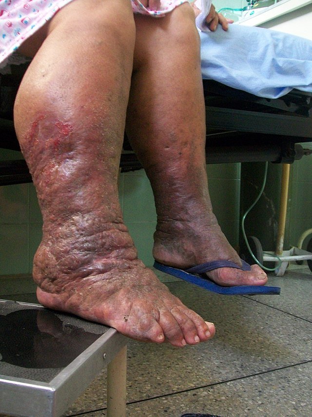
3. Advanced (Elephantiasis) Stage
Massive swelling with gross limb deformity
Skin changes:
Hyperkeratosis (thick, rough skin)
Wart-like papules or nodules
Lichenification (thickening from chronic irritation)
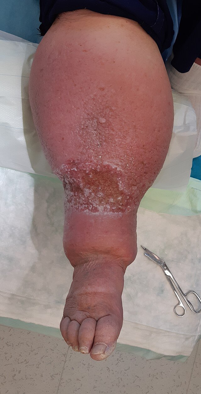
Increased infection risk:
Recurrent cellulitis or lymphangitis
May worsen oedema and accelerate fibrosis
4. Lymphoedema in Specific Conditions
Lymphatic Filariasis:
Often affects lower limbs, scrotum (hydrocele), vulva
May be bilateral but asymmetric
Chronic exposure leads to elephantiasis
Post-surgical/Radiation:
Affects arm (after breast surgery) or leg (after pelvic surgery)
Associated Symptoms
Limb heaviness or tightness
Skin tightness or discomfort
Reduced range of motion
Cosmetic concerns, anxiety, or social stigma
Investigations
1. Clinical Diagnosis First
Lymphoedema is largely a clinical diagnosis based on:
History (onset, location, precipitating events like surgery, travel)
Physical signs (non-pitting oedema, skin thickening, Stemmer’s sign)
However, investigations are needed to:
Confirm the diagnosis
Identify the underlying cause
Rule out alternative or coexisting pathology
2. Laboratory Investigations
| Test | Purpose |
|---|---|
| Full blood count | May show eosinophilia in parasitic infection (e.g. filariasis) |
| Filarial antigen card test | Detects Wuchereria bancrofti antigens (used any time of day) |
| Microfilariae in blood film | Best seen in night blood samples (10 pm–2 am) |
| Serology (ELISA/IFA) | Confirms filarial infection when microfilariae are absent |
| Urine examination | In suspected chyluria (milky urine, lymph leakage) |
3. Imaging Studies
a. Doppler Ultrasound
To exclude venous obstruction or thrombosis
Commonly used in leg swelling to rule out deep vein thrombosis (DVT)
b. Lymphoscintigraphy
Gold standard for assessing lymphatic function
Uses radiolabelled tracer to map lymph flow
Confirms delayed or absent lymph drainage
c. MRI / CT
Used to detect:
Soft tissue changes
Lymph node enlargement
Tumour compression or structural cause
d. Ultrasound of scrotum or limb
Can identify fluid collections, hydrocele, or adult worms (in filariasis)
4. Special Tests in Suspected Filariasis
PCR for filarial DNA – highly sensitive
Ultrasound of lymphatics – may show motile adult worms (“filarial dance sign”)
Chyle test – if urine is milky (chyluria)
5. When to Biopsy
Atypical features or unclear diagnosis
Rule out malignancy (lymphoma, metastatic cancer) if:
Rapid progression
Hard, fixed mass
No identifiable cause
Treatment
1. General Principles of Management
a. Limb Care and Skin Hygiene
Daily washing and drying of the limb
Moisturisation to prevent skin cracking
Prompt treatment of minor injuries to prevent cellulitis
Avoid tight clothing or trauma to the limb
b. Compression Therapy
Compression bandaging or garments (e.g. stockings, sleeves)
Promotes lymph drainage and prevents accumulation
Most effective in early or moderate disease
c. Limb Elevation and Exercise
Elevate limb during rest or sleep
Active limb exercises improve lymphatic flow
Avoid prolonged standing or dependency
2. Management of Secondary Causes
a. Lymphatic Filariasis
Diethylcarbamazine (DEC): kills microfilariae and adult worms
2 mg/kg TID for 12 days
Doxycycline: 200 mg/day for 4–8 weeks to target Wolbachia bacteria
Albendazole ± ivermectin: may be used in combination therapy
Mass Drug Administration (MDA): public health strategy to prevent transmission
b. Post-Surgical or Radiation Lymphoedema
Begin preventive limb care early after cancer treatment
Monitor for early swelling and start compression therapy promptly
3. Management of Complications
a. Recurrent Cellulitis
Prompt antibiotics for acute episodes (e.g. flucloxacillin, co-amoxiclav)
Prophylactic penicillin in patients with frequent cellulitis
b. Severe or Disfiguring Lymphoedema (Elephantiasis)
Intensive compression and hygiene may reduce limb size
Surgical options:
Debulking procedures (rarely curative)
Reserved for refractory or disabling cases
4. Patient Education
Emphasise long-term care and self-management
Monitor for early signs of infection
Encourage adherence to compression and follow-up
Prognosis & Follow-up
1. Prognosis
The prognosis of lymphoedema depends on:
Underlying cause
Stage at diagnosis
Timeliness of intervention
Patient adherence to lifelong care
Favourable outcomes
Early-stage or post-infective lymphoedema with good skin care and compression
Effective antiparasitic treatment in filarial cases
No secondary infections or complications
Poor outcomes
Late presentation with irreversible fibrosis and skin thickening
Recurrent bacterial cellulitis → further lymphatic damage
Neglect of skin care and compression
Elephantiasis: disfiguring, disabling, irreversible
2. Follow-up Goals
a. Monitor Disease Progression
Assess limb size, skin changes, and mobility at regular intervals
Watch for signs of worsening oedema or fibrosis
b. Prevent and Manage Infections
Educate on early signs of cellulitis: redness, pain, fever
Provide antibiotic prophylaxis if ≥2 infections/year
c. Support Adherence
Encourage long-term use of compression garments
Reinforce daily hygiene routines
d. Re-evaluate Secondary Causes
In suspected malignancy or TB, monitor response to treatment
For filariasis, assess efficacy of antiparasitic therapy and repeat testing if needed
3. Patient Education
Explain that lymphoedema is chronic but manageable
Highlight importance of:
Skin integrity
Compression use
Prompt response to infections
Disease-Specific Presentations
1. Lymphatic Filariasis
Most common global cause of lymphoedema
Caused by Wuchereria bancrofti, Brugia malayi
Chronic lymphatic obstruction due to adult worms and inflammation
Clinical features:
Progressive, non-pitting oedema of legs, scrotum, vulva, or breasts
Hydrocele in men
May be unilateral or asymmetric
Advanced cases develop elephantiasis: massive limb deformity and thickened, nodular skin
2. Post-Cancer Treatment (Surgical or Radiotherapy-Induced)
Common after:
Mastectomy or axillary dissection → arm lymphoedema
Pelvic cancer surgery → lower limb lymphoedema
Caused by lymphatic disruption or fibrosis
Onset may be delayed (weeks to months post-surgery)
Worsened by infection, obesity, or immobility
3. Tuberculous Lymphadenitis
Lymph node destruction and fibrosis of draining lymphatics
Seen in cervical or supraclavicular regions
May lead to localized lymphoedema of the face or neck
Sinus tract formation and matted nodes common
4. Congenital (Primary) Lymphoedema
Due to developmental abnormalities in lymphatic vessels
Subtypes:
Milroy disease: present at birth, often bilateral lower limb involvement
Meige disease: onset at puberty
Lymphoedema tarda: onset >35 years
May be familial or sporadic
5. Chronic Venous Insufficiency (Phlebolymphoedema)
Prolonged venous hypertension overwhelms lymphatic return
Occurs in older adults, often with varicose veins or DVT history
Mixed venous and lymphatic oedema in lower limbs
Often bilateral, worse at end of the day
6. Malignancy
Tumour compresses or invades lymphatics
Common in advanced cancers (e.g. pelvic, breast, lymphoma)
May present as:
Rapidly progressing oedema
Often unilateral and hard in consistency
Requires urgent imaging to assess for obstruction
7. Obesity-Associated Lymphoedema
Large body mass increases pressure on lymphatics
Skin folds promote infection and mechanical blockage
Affects lower limbs, may be confused with lipedema (fat deposition without oedema)