Leukoplakia
content of this page
1- Introduction
2- Clinical Features & Examination Tips
3- Investigations & Interpretation
4- Pathophysiology
5- Symptoms
6- Treatment
Introduction
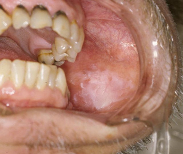
Leukoplakia refers to a persistent white patch in the oral mucosa that cannot be wiped off and cannot be classified as another condition. It is a clinical descriptor, not a diagnosis, but it is considered a potentially pre-malignant lesion, particularly if it persists despite removing local irritants.

Clinical Features & Examination Tips
-Suspect leukoplakia if you see:
A solitary white patch in the mouth that persists
Not removable by scraping (unlike candidiasis)
Often located on the tongue, buccal mucosa, or floor of mouth
-Examination tips:
Always examine dentures, dental fillings, and for chronic trauma
Check for cervical lymphadenopathy
Look for associated red patches (erythroplakia – higher risk)
Investigations & Interpretation
-First step: Rule out local causes (trauma, infection).
-Persistent patches >2 weeks → biopsy is mandatory.
Biopsy helps to assess:
Presence of dysplasia
Risk of malignant transformation
Pathophysiology
Leukoplakia is believed to result from chronic epithelial irritation, leading to hyperkeratosis (thickened keratin layer). In some cases, dysplasia or early squamous carcinoma develops. Risk factors include:
Tobacco use (smoking and smokeless forms)
Alcohol consumption
Chronic mechanical irritation
Symptoms
Usually asymptomatic, but may be:
Noticed incidentally by a dentist or clinician
Associated with mild discomfort or burning
In advanced cases, may ulcerate or harden (suggests malignancy)
Treatment
–Remove risk factors:
Stop tobacco and alcohol
Address dental trauma or irritation
-Monitor low-risk lesions
–Biopsy suspicious or persistent lesions
–Excision if:
Lesion shows dysplasia
Located in high-risk sites (floor of mouth, tongue)
Rapid growth or surface ulceration occurs
-Regular follow-up is essential due to malignant potential.