Coal Workers Pneumoconiosis (Anthracosilicosis)
content of this page
1- Definition & Types
2- Causes (Aetiology)
3- Pathophysiology
4- Clinical Features & Examination
5- Investigations
6- Management
7- Complications
8- Core Concepts
Definition & Types
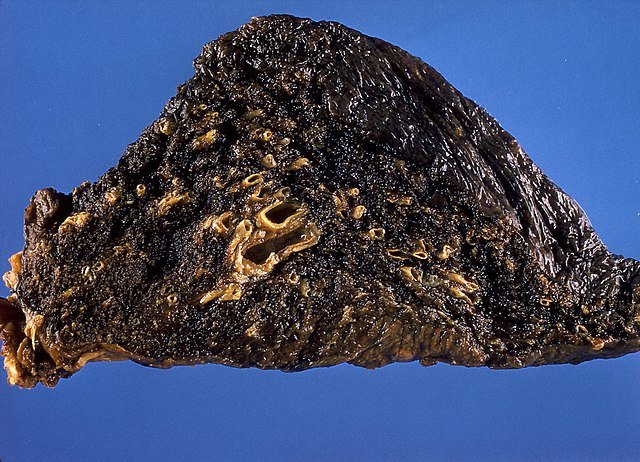
Coal Workers’ Pneumoconiosis is a chronic interstitial lung disease caused by long-term inhalation of coal dust, leading to fibrosis and structural lung damage.
Types:
Simple CWP (SCWP):
Characterized by small nodules in the upper lung zones.
Usually asymptomatic or mildly symptomatic.
Lung function often preserved.
Complicated CWP / Progressive Massive Fibrosis (PMF):
Coalescence of nodules into large fibrotic masses.
Leads to progressive dyspnoea, cough, and hypoxaemia.
Often continues progressing even after dust exposure ends.
Caplan’s Syndrome:
CWP + rheumatoid arthritis
Multiple rounded nodules (0.5–5 cm) with necrotic centers and inflammatory cells
Immune-mediated process

Causes (Aetiology)
Chronic exposure to inhaled coal dust in:
Underground mining
Coal processing
Dust contains carbon, silica, and other minerals
Disease severity correlates with:
Duration and intensity of exposure
Presence of coexisting conditions (e.g. RA)
Pathophysiology
Coal dust is inhaled and deposited in alveoli.
Alveolar macrophages engulf dust particles.
Accumulated dust and immune response → macule formation and fibrosis.
Ongoing inflammation leads to:
Nodules (in SCWP)
Large fibrotic masses (in PMF)
Autoimmune nodules in Caplan’s syndrome

Clinical Features & Examination
Symptoms:
SCWP: often asymptomatic or mild cough
PMF:
Persistent cough
Black sputum (melanoptysis)
Breathlessness
Weight loss and fatigue
Signs:
May include reduced breath sounds, fine crepitations, and cyanosis
No clubbing, unless complicated or with RA
Investigations
Chest X-ray:
SCWP: small nodular opacities in upper lobes
PMF: large masses (may cavitate), mimicking TB or cancer
High-resolution CT (HRCT):
Defines mass size, location, and cavitation
Pulmonary Function Tests:
Normal in SCWP
Restrictive ± obstructive in PMF
Autoimmune screen if Caplan’s suspected
TB testing to exclude differential
Management
No specific cure—management is supportive and preventative
Remove exposure: STOP coal dust inhalation
Vaccinations: influenza and pneumococcal
Treat infections promptly
Inhaled bronchodilators if airway obstruction
Oxygen therapy if hypoxaemic
Pulmonary rehabilitation
Monitor for TB, cancer, and RA symptoms
Complications
Progressive massive fibrosis (PMF)
Respiratory failure
Cor pulmonale (right heart failure)
Chronic bronchitis/emphysema overlap
Caplan’s syndrome (if RA is present)
Tuberculosis mimicry (must exclude)
Core Concepts
Fibrosis can progress even after exposure ends
Caplan’s = RA + CWP + nodules → think autoimmune
Always exclude TB in cavitating upper lobe lesions
Chest X-ray and CT are essential for staging
Prevention via dust control is key to public health
Lung function may remain intact in SCWP—don’t overdiagnose!