Colles’ Fracture
content of this page
1- Introduction
2- Anatomical Overview
3- Treatment
4- purposes
Introduction
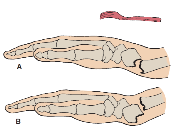
A Colles’ fracture is a break in the distal radius, the larger bone in the forearm, near the wrist. It typically occurs about an inch from the end of the radius, resulting in an upward and backward displacement of the bone, creating a “dinner fork” deformity.. Named after Abraham Colles, who first described it in 1814

Anatomical Overview
This fracture typically happens about an inch from the end of the radius and is characterized by the broken piece of the radius being displaced upward and backward. Often, the fracture may extend into the wrist joint, affecting its movement, and sometimes the ulnar styloid process, the tip of the ulna (the smaller bone in the forearm), may also be fractured. Soft tissues, including ligaments and tendons around the wrist, can be stretched or torn due to the displacement, and the median nerve, which runs through the carpal tunnel, may be compressed, leading to numbness or tingling in the fingers.

Treatment
The most common surgical procedure is open reduction and internal fixation (ORIF), which involves making an incision over the fractured area to directly access the broken bones. The bones are then realigned (reduced) into their correct position, and internal fixation devices such as plates, screws, or pins are used to hold the bones in place. ORIF is typically indicated for fractures with significant displacement or comminution (bone shattering) that cannot be adequately stabilized with a cast or splint alone
Purposes
The purpose of surgical intervention for a Colles’ fracture is to restore the fractured bones to their correct anatomical alignment and provide stable fixation to promote optimal healing and functional recovery.