Pterion Injury
content of this page
1- Introduction
2- Anatomical Overview
3- Causes
4- Treatment
Introduction
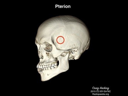
The pterion is a critical anatomical landmark on the lateral aspect of the skull where several bones converge: the frontal, parietal, temporal, and sphenoid bones. This region is vulnerable to injury due to its thin bone structure and proximity to major blood vessels, making pterion injuries significant in clinical practice.

Anatomical Overview
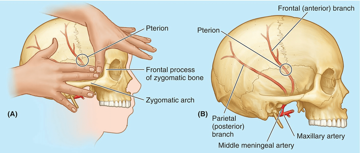
Fracture of the pterion can be life threatening because it overlies the frontal branches of the middle meningeal vessels, which lie in grooves on the internal aspect of the lateral wall of the calvaria. The pterion is two fingers’ breadth superior to the zygomatic arch and a thumb’s breadth posterior to the frontal process of the zygomatic bone. A hard blow to the side of the head (e.g., during boxing) may fracture the thin bones forming the pterion, producing a rupture of the frontal branch of the middle meningeal artery or vein crossing the pterion. The resulting hematoma exerts pressure on the underlying cerebral cortex. An untreated middle meningeal vessel hemorrhage may cause death in a few hours.

Causes
Blunt Trauma:
- Falls, motor vehicle accidents, assaults, and sports injuries can all lead to blunt force trauma to the head.
- Impact to the lateral aspect of the skull, where the pterion is located, can cause a fracture due to direct trauma.
Penetrating Trauma:
- Penetrating injuries such as gunshot wounds, stab wounds, or other sharp objects can directly impact the skull and cause fractures at the pterion.
- These injuries can be associated with significant morbidity and mortality due to potential damage to underlying structures and risk of intracranial bleeding.
Occupational Accidents:
- Individuals working in construction, mining, or other industries where head injuries are more common may sustain fractures of the pterion due to accidents involving falling objects or machinery.
Sports Injuries:
- High-impact sports such as football, hockey, or boxing can result in head trauma that may lead to fractures of the pterion.
- Falls or collisions during these activities can generate sufficient force to cause skull fractures in vulnerable areas like the pterion.
Child Abuse:
- In infants and young children, physical abuse can result in skull fractures, including those involving the pterion.
- Non-accidental trauma should always be considered in cases of pediatric head injuries.
Surgical Procedures:
- During surgical interventions involving the skull, inadvertent trauma or manipulation of the pterion area can lead to fractures.
- This is rare but can occur during procedures such as craniotomies or surgical access to the temporal or frontal regions of the skull.
Treatment
Initial Assessment and Stabilization:
- Airway, Breathing, Circulation (ABCs): Ensure that the patient’s airway is clear, breathing is adequate, and circulation is stable.
- Neurological Assessment: Perform a thorough neurological examination to assess for any signs of neurological deficits or changes in mental status.
Imaging Studies:
- CT Scan: Obtain a computed tomography (CT) scan of the head to evaluate the extent and characteristics of the fracture.
- CT imaging helps assess for associated injuries, such as intracranial hemorrhage (e.g., epidural hematoma) or other skull fractures.
Management of Associated Injuries:
- If there is evidence of intracranial bleeding, particularly from injury to the middle meningeal artery near the pterion, urgent neurosurgical consultation may be required.
- Surgical intervention, such as evacuation of hematoma or repair of vascular injury, may be necessary to prevent neurological deterioration.
Non-Surgical Management:
- Stable fractures without significant intracranial complications may be managed conservatively.
- Pain management and monitoring for signs of increased intracranial pressure are essential during the initial observation period.
Surgical Intervention:
- Craniotomy: In cases of severe trauma with significant intracranial hemorrhage or underlying brain injury, a craniotomy may be performed to evacuate hematomas, relieve pressure, and repair skull fractures.
- Repair of Skull Defects: If the fracture involves a significant defect or disruption of the skull, surgical repair may be considered to prevent complications such as cerebrospinal fluid (CSF) leaks or infection.
Monitoring and Follow-Up:
- Close monitoring of neurological status, vital signs, and imaging studies is crucial during the initial post-injury period.
- Follow-up visits with neurosurgery and/or trauma teams are necessary to assess recovery, manage complications, and ensure optimal healing of the fracture.
Rehabilitation and Long-Term Care:
- Depending on the severity of associated injuries and neurological deficits, rehabilitation services may be needed to facilitate recovery and optimize functional outcomes.
- Long-term management may include follow-up imaging studies, neurological assessments, and ongoing care for any residual symptoms or complications.