Cervical Rib
content of this page
1- Introduction
2- Anatomical Overview
3- Causes
4- Treatment
Introduction

Anatomical Overview
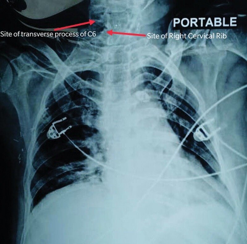
A cervical rib is a relatively common anomaly. In 1–2% of people, the developmental costal element of C7, which normally becomes a small part of the transverse process that lies anterior to the foramen transversarium, becomes abnormally enlarged. This structure may vary in size from a small protuberance to a complete rib that occurs bilaterally about 60% of the time.
The supernumerary (extra) rib or a fibrous connection extending from its tip to the first thoracic rib may elevate and place pressure on structures that emerge from the superior thoracic aperture, notably the subclavian artery or inferior trunk of the brachial plexus, and may cause thoracic outlet syndrome.

Causes
Genetic Factors: Genetic predisposition may play a role in the formation of cervical ribs. There may be familial patterns or genetic mutations that increase the likelihood of developing cervical ribs.
Embryological Development: During fetal development, the formation of ribs occurs through a complex process involving the differentiation of mesenchymal cells and ossification centers. Aberrations in this process can lead to the formation of an extra rib at the cervical level.
Hox Genes: Hox genes are a family of genes that play a crucial role in the patterning of the body during embryonic development. Mutations or dysregulation of Hox genes may contribute to the development of cervical ribs.
Environmental Factors: While the exact environmental factors contributing to cervical rib formation are not fully understood, certain environmental influences during pregnancy may affect embryonic development and increase the risk of congenital anomalies.
Trauma: In rare cases, trauma or injury to the neck area during fetal development or early childhood may disrupt normal bone development and contribute to the formation of cervical ribs. However, trauma is not a common cause of cervical ribs.
Idiopathic Causes: In some instances, the exact cause of cervical rib formation may not be identified, and the anomaly is considered idiopathic.
Treatment
Conservative Management:
- Physical Therapy: Physical therapy focuses on strengthening and stretching exercises to improve posture, alleviate muscle tension, and relieve pressure on neurovascular structures in the thoracic outlet region.
- Ergonomic Modifications: Advising patients on ergonomic adjustments in daily activities, such as avoiding prolonged overhead activities and maintaining proper posture, can help reduce symptoms associated with TOS.
- Pain Management: Nonsteroidal anti-inflammatory drugs (NSAIDs), muscle relaxants, or neuropathic pain medications may be prescribed to alleviate pain and discomfort associated with TOS.
Interventional Procedures:
- Nerve Blocks: Local anesthetic injections or nerve blocks may be administered to temporarily block nerve signals and provide relief from pain associated with TOS.
- Botulinum Toxin Injections: In some cases, botulinum toxin injections into hypertonic muscles in the neck and shoulder region may help alleviate muscle spasms and reduce compression on neurovascular structures.
Surgical Options:
- First Rib Resection: Surgical removal of the cervical rib, along with the first rib if necessary, is often considered when conservative measures fail to provide adequate relief or when there are significant neurological or vascular complications associated with TOS. This procedure aims to decompress the thoracic outlet and relieve pressure on affected structures.
- Scalenectomy: In cases where muscle hypertrophy or fibrosis contributes to thoracic outlet compression, surgical excision of the scalene muscles (scalenectomy) may be performed in conjunction with first rib resection to widen the thoracic outlet.
- Thoracic Outlet Decompression: Various surgical techniques, including transaxillary, supraclavicular, or infraclavicular approaches, may be employed to decompress the thoracic outlet and alleviate symptoms of TOS.
Postoperative Rehabilitation:
- Following surgical intervention, patients may undergo postoperative rehabilitation, including physical therapy and occupational therapy, to regain strength, range of motion, and functional abilities in the affected upper limb.