Dupuytren’s Contracture
content of this page
1- Introduction
2- Anatomical Overview
3- Treatment
Introduction

Anatomical Overview
The palmar aponeurosis is a crucial structure within the palm of the hand, playing a key role in hand function and mechanics. Here’s an overview of its anatomy, function, and clinical significance:
Anatomy of the Palmar Aponeurosis
Location: The palmar aponeurosis is a thick, triangular fibrous sheet located superficially in the center of the palm, extending from the distal end of the flexor retinaculum to the bases of the fingers.
Structure:
- Proximal Attachment: It is continuous with the palmaris longus tendon (when present) and the flexor retinaculum.
- Distal Attachment: It divides into four slips that extend into the fingers. Each slip further divides into two bands that merge with the fibrous digital sheaths and the deep transverse metacarpal ligaments.
Composition: The aponeurosis is made of dense connective tissue, providing strength and protection to the underlying structures.
Function of the Palmar Aponeurosis
- Protection: It protects the underlying muscles, nerves, and blood vessels in the palm.
- Support: It provides structural support to the palm, maintaining its shape and allowing efficient force transmission.
- Attachment: It serves as an attachment site for the palmaris longus tendon (if present) and helps anchor the skin, enhancing grip and preventing slippage during object manipulation.
Treatment
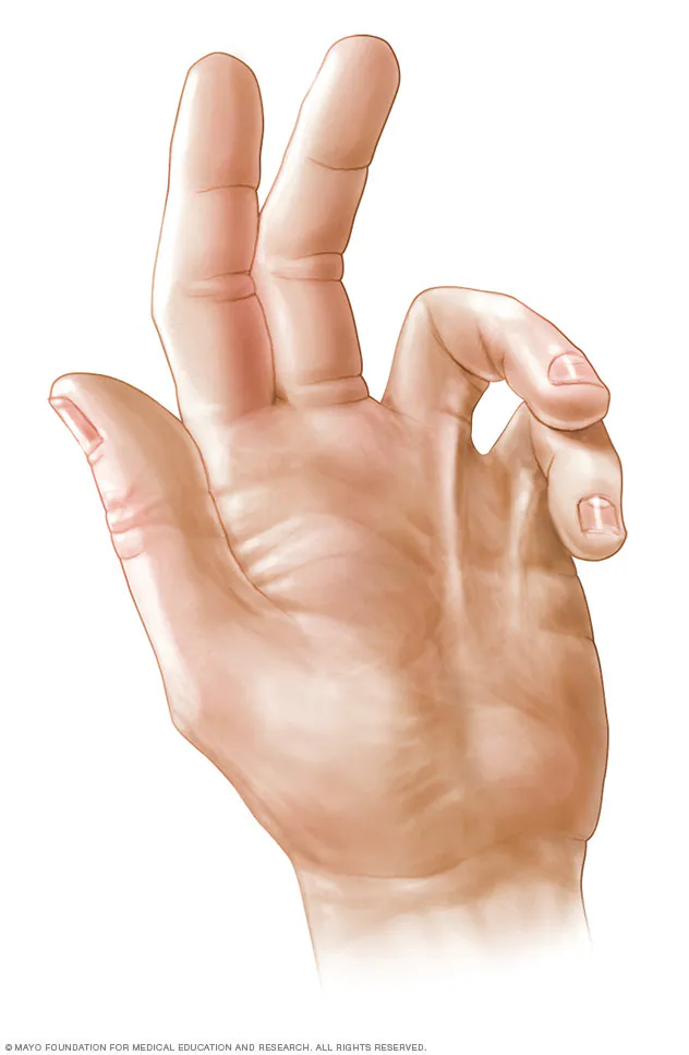
Surgical division of the fibrous bands followed by physiotherapy to the hand is the usual form of treatment. The alternative treatment of injection of the enzyme collagenase into the contracted bands of fibrous tissue has been shown to significantly reduce the contractures and improve mobility.