Studying the diffraction using laser beam by: a slit and a hole
Content of this page :
1. Introduction of the experiment.
2. Aim of the experiment.
3. Tools of the experiment.
4. Steps and methods of the experiment.
5. Parameter of the experiment.
6. Medical application and advantages of the experiment.
1. Introduction of the experiment:
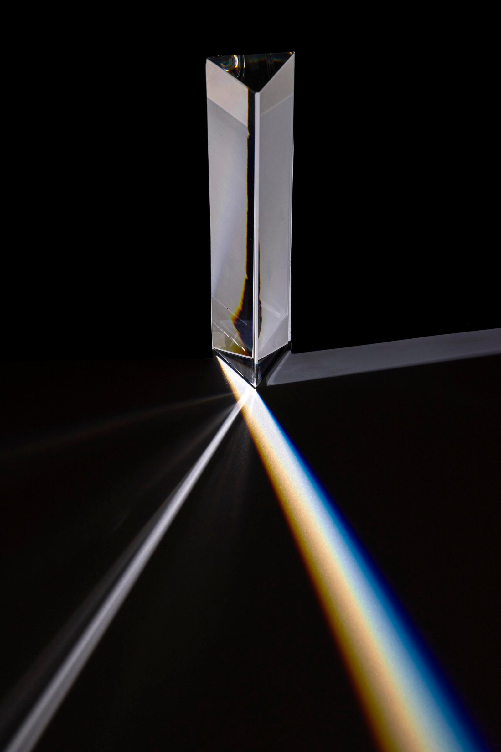
The term diffraction refers to phenomena in which light or other radiation exhibits departures from the straight line propagation predicted by the simplified model of geometrical optics. Analysis of diffraction phenomena always requires the more complete description given by the wave picture of light. The calculation of the principle characteristics of simple diffraction patterns are usually based on Huygens’ principle.
2. Aim of the experiment:
-The aim of the experiment studying diffraction using a laser beam through a slit and a hole is to investigate and demonstrate the principles of diffraction of light.

3-Tools of The experiment
- Laser Source
- Optical Bench:
- Slit and Aperture Components
- Screen or Detector
- Measurement Tools
- Additional Components
- Safety Equipment
- Computer and Software (Optional)
4. Steps and methods of the experiment:
Aim the laser beam at one of the single slit apertures and measure the position of the intensity minimum in the diffraction pattern. Compute Xo L. then obtain the the corresponding values of: (tan0) corresponding values of: (sin0), and plot a graph of: (sin0) as a fraction of: (n). Using this graph, together with equation one, determine the slit width (d). Replace the single slit with a circular hole of diameter (d), then obtain the diffraction pattern which is a general bright circle of light surrounded by concentric alternate dark and light rings. The lines joining the hole to the first dark ring forma cone, and it is found that the semi-
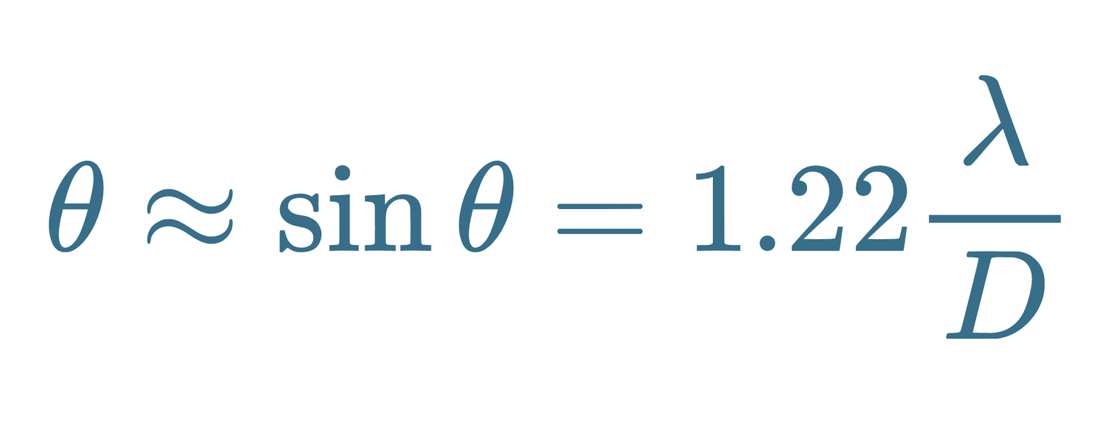
angle of this cone is (0), where:
Compute (0), and using the known value of (2) for the He-Ne laser, determine the diameter (d) of the circular hole.
5-Parameter of The Experiment:
Complete destructive interference occurs at points for which:

Between these points of zero intensity are other regions of maximum intensity, but not as bright as the general maximum, (why?).
it is found that the semi-angle of this cone is (0), where:

Compute (0), and using the known value of (2) for the He-Ne laser, determine the diameter (d) of the circular hole.
6-Medical application
Studying diffraction using laser beams through slits and holes has several potential medical applications, particularly in the field of optics and medical diagnostics. Here are a few examples:
Optical Coherence Tomography (OCT):
- Application: OCT is a non-invasive imaging technique used in ophthalmology and other medical fields to obtain high-resolution cross-sectional images of biological tissues.
- Use of Diffraction: Diffraction principles, including those studied with slits and holes, are fundamental in understanding and optimizing the performance of OCT systems. They help in designing the optical components such as beam splitters and mirrors to achieve the desired beam characteristics and interference patterns crucial for image resolution.
Laser Scalpel and Surgery:
- Application: High-precision laser surgery techniques rely on controlling the laser beam’s properties to accurately cut or ablate tissue without damaging surrounding areas.
- Use of Diffraction: Understanding diffraction helps in designing laser delivery systems with precise beam shaping and focusing capabilities. Diffraction principles can optimize the laser’s spatial coherence and divergence characteristics, which are critical for surgical accuracy.
Flow Cytometry:
- Application: Flow cytometry is a technique used in medical diagnostics and research to analyze characteristics of cells and particles in fluid suspension.
- Use of Diffraction: Diffraction techniques can be applied to develop advanced flow cytometry systems with improved light scattering and detection capabilities. Understanding diffraction helps in optimizing the design of light sources, detectors, and optical filters to enhance sensitivity and resolution in detecting cellular characteristics.
Diagnostic Imaging Techniques:
- Application: Various imaging techniques in medicine, such as confocal microscopy and fluorescence imaging, rely on precise control of light beams and their interaction with biological samples.
- Use of Diffraction: Diffraction studies assist in optimizing these imaging systems to achieve high-resolution and high-contrast images. Understanding diffraction helps in designing optical components like lenses, mirrors, and filters to improve light collection efficiency and minimize artifacts.
Laser Doppler Blood Flowmetry:
- Application: Laser Doppler flowmetry is used to measure blood flow in tissues, which is important in monitoring conditions like peripheral vascular disease and wound healing.
- Use of Diffraction: Diffraction principles are crucial in designing laser beams that can penetrate tissues effectively and generate reliable Doppler signals. Optimization of laser beam characteristics through diffraction studies contributes to improving the accuracy and sensitivity of blood flow measurements.
