

Clinical Subject Page
Pulmonary Valve Disease (Stenosis & Regurgitation)
Also called
ICD-10
Specialty
Onset
Reviewed
-
OverviewOverview
-
Etiology & Risk FactorsEtiology & Risk Factors
-
PathophysiologyPathophysiology
-
Clinical PresentationClinical Presentation
-
History TakingHistory Taking
-
Pulmonary Stenosis Vs Pulmonary RegurgitationPulmonary Stenosis Vs Pulmonary Regurgitation
-
InvestigationsInvestigations
-
DiagnosisDiagnosis
-
ManagementManagement
-
ComplicationsComplications
-
PrognosisPrognosis
-
Key Points / Clinical PearlsKey Points / Clinical Pearls
Overview
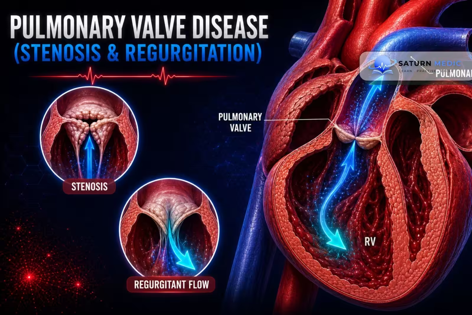
Pulmonary valve disease affects the pulmonary valve, which is located between the right ventricle (RV) and the pulmonary artery. It includes:
- Pulmonary Stenosis (PS): Narrowing of the pulmonary valve that obstructs blood flow from the right ventricle to the pulmonary artery during systole. It is usually congenital.
- Pulmonary Regurgitation (PR): Failure of the pulmonary valve to close properly, causing backflow of blood from the pulmonary artery into the right ventricle during diastole.
Etiology & Risk Factors
1. Pulmonary Stenosis (PS)
Most Common Cause
Congenital pulmonary valve stenosis (most cases)
Other Causes (Rare)
Rheumatic fever
Carcinoid heart disease
2. Pulmonary Regurgitation (PR)
Common Causes
Pulmonary hypertension (most common cause of significant PR)
Pulmonary valve damage after balloon valvuloplasty or valve surgery
Congenital heart disease
Infective endocarditis
Carcinoid heart disease
General Risk Factors (Pulmonary Valve Disease)
Congenital heart disease
Noonan syndrome (associated with pulmonary stenosis)
Rheumatic fever
Carcinoid syndrome/heart disease
Pulmonary hypertension
Previous pulmonary valve intervention or surgery
Infective endocarditis
Pathophysiology
1. Pulmonary Stenosis (PS)
Narrowing of the pulmonary valve obstructs blood flow from the right ventricle (RV) to the pulmonary artery during systole.
This causes:
Right ventricular outflow obstruction
Pressure overload of the RV
Right ventricular hypertrophy (RVH)
If severe, right-sided heart failure
Simple Flow:
Pulmonary valve narrowing → RV outflow obstruction → RV pressure overload → RV hypertrophy → Right-sided heart failure
2. Pulmonary Regurgitation (PR)
Pulmonary valve fails to close properly, allowing blood to flow back from the pulmonary artery into the RV during diastole.
This causes:
Right ventricular volume overload
Progressive RV dilation
Eventually right-sided heart failure if severe
Simple Flow:
Pulmonary valve incompetence → Blood leaks back into RV → RV volume overload → RV dilation → Right-sided heart failure
Clinical Presentation
-Pulmonary Stenosis (PS)
Symptoms
May be asymptomatic if mild.
Exercise intolerance
Symptoms of heart failure in more severe disease.
Physical Examination
Crescendo–decrescendo systolic murmur heard best at the 2nd left intercostal space (left upper sternal border).
Wide splitting of S₂.
Pulmonary Regurgitation (PR)
Symptoms
Often asymptomatic if mild.
If severe:
Fatigue
Exercise intolerance
Symptoms of right-sided heart failure
Physical Examination
Early diastolic decrescendo murmur heard best at the 2nd left intercostal space.
Graham Steell murmur may be present.
History Taking
Symptoms:
“Do you get short of breath during exercise or daily activities?”
“Do you feel tired more easily than before?”
“Do you have chest pain?”
“Do you feel your heart racing or skipping beats (palpitations)?”
“Have you ever fainted or felt dizzy?”
Symptoms of Right-Sided Heart Failure
“Do you have swelling in your legs or ankles?”
“Have you noticed abdominal swelling or weight gain due to fluid?”
Risk Factors
“Were you born with a heart defect?”
“Do you have Noonan syndrome?”
“Have you had rheumatic fever?”
“Have you ever had infective endocarditis?”
“Have you had previous pulmonary valve surgery or balloon valvuloplasty?”
“Have you been told you have pulmonary hypertension?”
Pulmonary Stenosis Vs Pulmonary Regurgitation
Pulmonary Stenosis vs Pulmonary Regurgitation
| Feature | Pulmonary Stenosis | Pulmonary Regurgitation |
|---|---|---|
| Murmur | Systolic ejection murmur, crescendo-decrescendo, upper left sternal border, radiates to back; widely split S2 with soft P2 | Early diastolic decrescendo murmur (Graham Steell if 2° to pulmonary hypertension), upper left sternal border |
| JVP findings | Prominent "a" wave if RV hypertrophy reduces compliance | Usually unremarkable unless severe RV failure develops; may see elevated JVP late |
| Pulse/other signs | RV heave; ejection click that decreases with inspiration (unusual right-sided finding) | RV heave if RV dilated; signs of underlying pulmonary hypertension |
| Key echo finding | Increased systolic gradient across pulmonary valve (peak velocity/gradient); doming valve; post-stenotic PA dilation | Color Doppler diastolic regurgitant jet; PR jet width/duration; RV dilation; paradoxical septal motion if severe |
| Right heart chamber effect | RV hypertrophy predominates (pressure overload); RV size may be normal early | RV dilation predominates (volume overload); progressive RV dysfunction if chronic severe |
| Common associations | Congenital heart disease, Noonan syndrome, tetralogy of Fallot, carcinoid (rare) | Pulmonary hypertension, post-TOF repair, post-balloon valvotomy, endocarditis (rare), carcinoid |
| Definitive treatment | Balloon valvuloplasty (first-line for valvular PS); surgical valvotomy/replacement if unsuitable | Treat underlying cause; pulmonary valve replacement (surgical or transcatheter) if severe with RV dilation/dysfunction or symptoms |
Investigations
Investigations (Ix) – Pulmonary Valve Disease (PS & PR)
1. Transthoracic Echocardiography (TTE) – Gold Standard
First-line and confirmatory test
Assesses:
Valve structure
Severity of pulmonary stenosis (PS) or pulmonary regurgitation (PR)
Right ventricular function
Pressure gradient across the pulmonary valve (for PS)
2. ECG
May be normal in mild PS.
May show right ventricular hypertrophy (RVH) in severe disease.
3. Chest X-ray
May show pulmonary artery dilatation.
4. Cardiac Catheterization
Performed in selected patients, especially before valve repair when indicated.
High-Yield Point
TTE is the gold-standard investigation for pulmonary valve disease.
Diagnosis
Diagnostic Approach
Suspect pulmonary valve disease based on:
Symptoms
Characteristic heart murmur on examination
Confirm the diagnosis with transthoracic echocardiography (TTE).
Gold-Standard Test
Transthoracic echocardiography (TTE)
Confirms the diagnosis.
Assesses the severity of pulmonary stenosis and valve function.
Supportive Investigations
ECG
May be normal in mild pulmonary stenosis.
May show right ventricular hypertrophy (RVH) in severe disease.
Chest X-ray
May show pulmonary artery dilatation.
Cardiac catheterization
Considered before repair in selected patients.
Management
1. Pulmonary Stenosis (PS)
Mild Disease
Regular follow-up with clinical assessment and echocardiography.
Symptomatic Moderate-to-Severe PS
Balloon pulmonary valvuloplasty (preferred treatment).
Pulmonary valve replacement if balloon valvuloplasty is not suitable or unsuccessful.
Critical Pulmonary Stenosis in Neonates
Maintain the patent ductus arteriosus (PDA) with prostaglandin E₁ (alprostadil) until definitive treatment can be performed.
2. Pulmonary Regurgitation (PR)
Treat the underlying cause (e.g., pulmonary hypertension).
Monitor with regular echocardiography.
Pulmonary valve replacement may be required in severe symptomatic disease or when there is significant right ventricular dysfunction.
Complications
- Right ventricular hypertrophy (RVH) (especially in pulmonary stenosis)
- Right ventricular dilation (especially in pulmonary regurgitation)
- Right-sided heart failure
- Exercise intolerance
- Progressive valve dysfunction if left untreated
Prognosis
- Mild pulmonary valve disease generally has a good prognosis and often requires only regular follow-up.
- Prognosis worsens with severe disease, development of right ventricular dysfunction, or heart failure.
- Early intervention (e.g., balloon valvuloplasty for pulmonary stenosis or valve replacement when indicated) improves outcomes.
Key Points / Clinical Pearls
- Pulmonary stenosis (PS) = narrowed pulmonary valve → right ventricular pressure overload.
- Pulmonary regurgitation (PR) = leaky pulmonary valve → right ventricular volume overload.
- Most PS cases are congenital.
- Gold-standard investigation: Transthoracic echocardiography (TTE).
- PS murmur: Crescendo–decrescendo systolic murmur at the 2nd left intercostal space.
- PR murmur: Early diastolic decrescendo (Graham Steell) murmur.
- Preferred treatment for symptomatic moderate-to-severe PS: Balloon pulmonary valvuloplasty.
- Both conditions can eventually lead to right-sided heart failure if severe or untreated.
- Heaton J, Horenstein MS, Kyriakopoulos C. National Center for Biotechnology Information (NIH). Pulmonary Stenosis, StatPearls.
- National Center for Biotechnology Information (NIH). Pulmonary Regurgitation, StatPearls.
- Pulmonary Valve Stenosis: From Diagnosis to Current Management Techniques and Future Prospects. PMC10320808.
- Bouzas B, Kilner PJ, Gatzoulis MA. Pulmonary Regurgitation: Not a Benign Lesion. Eur Heart J. 2005;26:433-439. PMC1994453.
- MedlinePlus, National Library of Medicine (NIH). Pulmonary Valve Stenosis: Medical Encyclopedia.