

Clinical Subject Page
Pulmonary Embolism (PE)
ICD-10
Specialty
Onset
Reviewed
June 2026
-
OverviewOverview
-
Etiology & Risk FactorsEtiology & Risk Factors
-
PathophysiologyPathophysiology
-
Clinical PresentationClinical Presentation
-
History TakingHistory Taking
-
ClassificationClassification
-
Investigations ApproachInvestigations Approach
-
DiagnosisDiagnosis
-
ManagementManagement
-
ComplicationsComplications
-
PrognosisPrognosis
-
Key Points / Clinical PearlsKey Points / Clinical Pearls
Overview
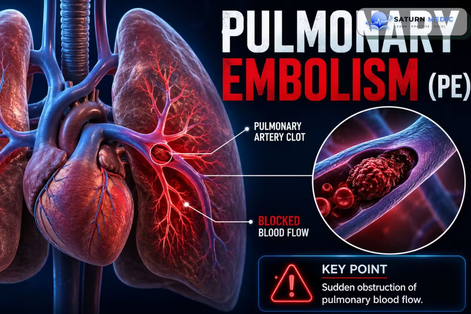
Pulmonary embolism (PE) is the obstruction of one or more pulmonary arteries, usually by a thrombus originating from a deep vein thrombosis (DVT), leading to impaired pulmonary blood flow and impaired gas exchange.
Etiology & Risk Factors
Etiology of Pulmonary Embolism (PE)
Deep vein thrombosis (most common)
Surgery (especially orthopedic)
Trauma
Malignancy
Pregnancy/postpartum
Prolonged immobilization
Inherited thrombophilia
Estrogen therapy (OCP/HRT)
Previous VTE
Risk Factors for Pulmonary Embolism (PE)
Previous DVT/PE
Active cancer
Recent surgery
Hospitalization
Long-distance travel
Obesity
Smoking
Heart failure
Pregnancy
Increasing age
Pathophysiology
- A blood clot forms in a deep vein of the leg or pelvis (DVT).
- The clot breaks off and travels through the inferior vena cava (IVC) to the pulmonary arteries.
- The clot blocks blood flow to part of the lung.
- This causes:
- Less oxygen enters the blood (hypoxemia) because blood cannot reach ventilated alveoli (V/Q mismatch).
- Chest pain and hemoptysis if lung tissue becomes infarcted.
- The blockage also increases pressure in the pulmonary arteries, making the right ventricle work harder.
- In a large (massive) PE, the right ventricle can fail, causing:
- Low blood pressure
- Shock
- Collapse
Flow Chart
DVT → Clot travels to lungs → Pulmonary artery blockage → V/Q mismatch → Hypoxemia + Increased right heart workload → Right ventricular failure (if severe) → Shock
Clinical Presentation
Symptoms
- Sudden dyspnea
- Pleuritic chest pain
- Cough
- Hemoptysis
- Syncope
- Palpitations
- Anxiety
Signs
- Tachypnea
- Tachycardia
- Hypoxemia
- Fever (low-grade)
- Elevated JVP (massive PE)
- Hypotension (massive PE)
- Signs of DVT
History Taking
-
Key Questions
- Sudden onset of shortness of breath?
- Chest pain (pleuritic)?
- Hemoptysis?
- Leg swelling or pain?
- Recent surgery or hospitalization?
- Long travel or immobilization?
- Previous DVT or PE?
- Cancer history?
- Pregnancy or OCP use?
Red Flags for Pulmonary Embolism (PE)
- Hypotension
- Syncope
- Severe hypoxemia
- Cardiac arrest
- Massive hemoptysis
Classification
Pulmonary Embolism · Classification
| Classification Basis | Categories |
|---|---|
| By Haemodynamic Risk (ESC) | High-risk (massive) Intermediate-risk (submassive) Low-risk |
| By Timing of Presentation | Acute (symptoms develop immediately) vs Chronic (CTEPH — persistent perfusion defects ≥3 months post-PE) |
| By Anatomical Location | Saddle embolus (straddles main PA bifurcation) — Lobar — Segmental — Subsegmental |
| By Source/Provoking Factor | Provoked (surgery, immobilisation, malignancy, hormones) vs Unprovoked (no identifiable transient risk factor — longer anticoagulation typically considered) |
| By Recurrence | First episode vs Recurrent PE — influences duration/intensity of anticoagulation |
Investigations Approach
Pulmonary Embolism · Investigations by Haemodynamic Status
Diagnosis
1. Clinical Assessment
Symptoms and signs
Assessment of risk factors
Estimate pretest probability using a clinical prediction rule (e.g., Wells score or Geneva score)
2. D-dimer
Use in patients with low or intermediate clinical probability
A negative D-dimer can effectively exclude PE in appropriate patients
3. CT Pulmonary Angiography (CTPA) — Gold Standard Imaging Test
First-line imaging test for most hemodynamically stable patients
Demonstrates filling defects within the pulmonary arteries
4. Ventilation–Perfusion (V/Q) Scan
Consider when:
CTPA is contraindicated
Severe contrast allergy
Pregnancy (selected patients)
Significant renal impairment
5. Compression Ultrasonography of the Legs
Detects deep vein thrombosis (DVT)
Supports the diagnosis when CTPA cannot be performed
6. Echocardiography
Useful in:
Hemodynamically unstable patients
Suspected massive Pulmonary Embolism (PE)
May show:
Right ventricular dilation or dysfunction
Management
Initial Management
Oxygen therapy
Hemodynamic stabilization
Analgesia
Anticoagulation
Low-molecular-weight heparin
Unfractionated heparin
Direct oral anticoagulants (Apixaban, Rivaroxaban)
Warfarin (selected patients)
Reperfusion Therapy
Systemic thrombolysis (massive Pulmonary Embolism PE)
Catheter-directed thrombolysis
Surgical embolectomy
Additional Therapy
Inferior vena cava (IVC) filter (if anticoagulation is contraindicated)
Complications
-
- Acute right ventricular failure
- Cardiogenic shock
- Cardiac arrest
- Recurrent Pulmonary Embolism (PE)
- Chronic thromboembolic pulmonary hypertension (CTEPH)
- Death
Prognosis
- Excellent with early diagnosis and anticoagulation.
- Massive Pulmonary Embolism (PE) carries a high mortality if untreated.
- Right ventricular dysfunction is the strongest predictor of poor outcome.
Key Points / Clinical Pearls
- Most Pulmonary Embolisms (PE) originate from lower-extremity DVT.
- CT pulmonary angiography is the diagnostic imaging modality of choice.
- Use the Wells score to assess pretest probability.
- Start anticoagulation promptly when PE is strongly suspected unless contraindicated.
- Hemodynamic instability indicates high-risk (massive) Pulmonary Embolism (PE) and may require thrombolysis or embolectomy.
- Chronic thromboembolic pulmonary hypertension is an important long-term complication.
- Vyas V, Sankari A, Goyal A. National Center for Biotechnology Information (NIH). Acute Pulmonary Embolism, StatPearls.
- Konstantinides SV, Meyer G, Becattini C, et al. 2019 ESC Guidelines for the Diagnosis and Management of Acute Pulmonary Embolism Developed in Collaboration With the European Respiratory Society (ERS). Eur Respir J. 2019;54:1901647. PMID: 31473594.
- Shafi I, Devarapally SR, Gupta N. National Center for Biotechnology Information (NIH). Catheter-Directed Thrombolysis of Pulmonary Embolism, StatPearls.
- MedlinePlus, National Library of Medicine (NIH). Pulmonary Embolism: Health Topic.
- National Center for Biotechnology Information (NIH). Deep Vein Thrombosis, StatPearls.