
Upper Limb Anatomy PDF
Explore the structure and function of the upper limb through detailed anatomy modules.
What's Covered in the Upper Limb Anatomy PDF
Welcome to the Saturn Medic upper limb anatomy PDF — a free, structured set of study notes covering the shoulder, arm, forearm, wrist, and hand. Each section is previewable directly on the site, with a full downloadable PDF available for offline study before labs, practicals, or exams. This upper limb anatomy PDF was built to give medical students a clear, consistent reference for one of the most heavily tested regions in early anatomy courses.
What’s Covered in the Upper Limb Anatomy PDF
The upper limb anatomy PDF is organized to match how the region is typically taught, moving through the major structural categories:
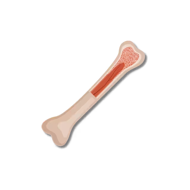
Osteology — the bones of the upper limb, from the clavicle and scapula through the humerus, radius, ulna, and the carpal, metacarpal, and phalangeal bones of the hand, with the landmarks and articulation points most often referenced in exams.
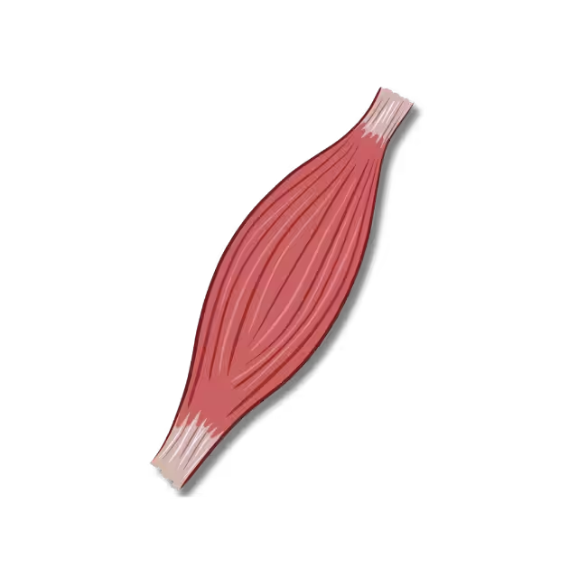
Myology — the muscles of the shoulder, arm, forearm, and hand, grouped by compartment and function, covering origin, insertion, action, and innervation for each.
Arthrology — the joints of the upper limb, including the shoulder, elbow, wrist, and hand joints, along with their supporting ligaments and typical ranges of motion.
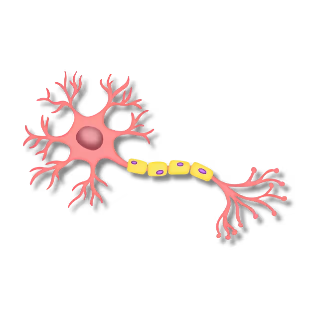
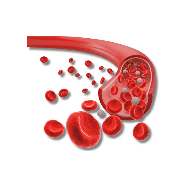
Neurovascular Structures — the nerves and vessels running through the upper limb, including the brachial plexus and its major branches, axillary contents, and the vascular supply from the subclavian artery through to the hand — consistently one of the highest-yield and most exam-relevant parts of upper limb anatomy.
Each section of the upper limb anatomy PDF follows the same consistent format, so once you’re comfortable with how one structural category is presented, the rest of the document feels immediately familiar.
Why This Upper Limb Anatomy PDF Helps
The upper limb is dense with clinically relevant detail packed into a relatively small area — brachial plexus injuries, axillary lymph node relationships, carpal tunnel syndrome, and compartment-specific muscle testing all come from structures covered in this region, which is exactly why it shows up so heavily in both anatomy and clinical exams. Having a single, well-organized upper limb anatomy PDF makes it easier to hold all of that detail together instead of piecing it back from scattered lecture slides or a heavy textbook chapter.
Our upper limb anatomy PDF content is styled after standard clinical anatomy references such as Snell’s Clinical Anatomy and Netter’s Atlas of Human Anatomy, with structural details cross-checked against interactive 3D tools like Anatomy 3D Atlas for accuracy. This keeps the terminology and structural relationships in this upper limb anatomy PDF consistent with what students already encounter in their coursework, rather than presenting a simplified or inconsistent version of the material.
These notes are built to complement hands-on lab work, not replace it. Reviewing the upper limb anatomy PDF before a cadaveric dissection or model-based lab session means you walk in already familiar with the major landmarks you’ll be asked to identify, which makes that lab time far more productive than trying to learn the structures for the first time under pressure.
How to Use This Upper Limb Anatomy PDF
Start with the structural category most relevant to what you’re currently studying — Osteology, Myology, Arthrology, or Neurovascular Structures — and preview it directly on the site, or download the full upper limb anatomy PDF for offline study or printing. Many students pair this upper limb anatomy PDF with the Upper Limb category in our anatomy question bank, using the notes to build understanding first and the MCQs to test recall afterward under exam-style time pressure — the combination tends to stick better than either resource used alone.
New content and updates continue to be added to the upper limb anatomy PDF as the broader anatomy lab library expands across all 8 body regions.