

Clinical Subject Page
Chronic Coronary Syndrome (CCS)
Commonly known as a heart attack — irreversible necrosis of myocardial tissue caused by prolonged ischemia, most often due to occlusion of a coronary artery.
Also called
ICD-10
Specialty
Onset
Reviewed
-
OverviewOverview
-
Etiology & Risk FactorsEtiology & Risk Factors
-
PathophysiologyPathophysiology
-
Clinical PresentationClinical Presentation
-
History TakingHistory Taking
-
Physical ExaminationPhysical Examination
-
InvestigationsInvestigations
-
DiagnosisDiagnosis
-
ManagementManagement
-
ComplicationsComplications
-
PrognosisPrognosis
-
Key Points / Clinical PearlsKey Points / Clinical Pearls
Overview
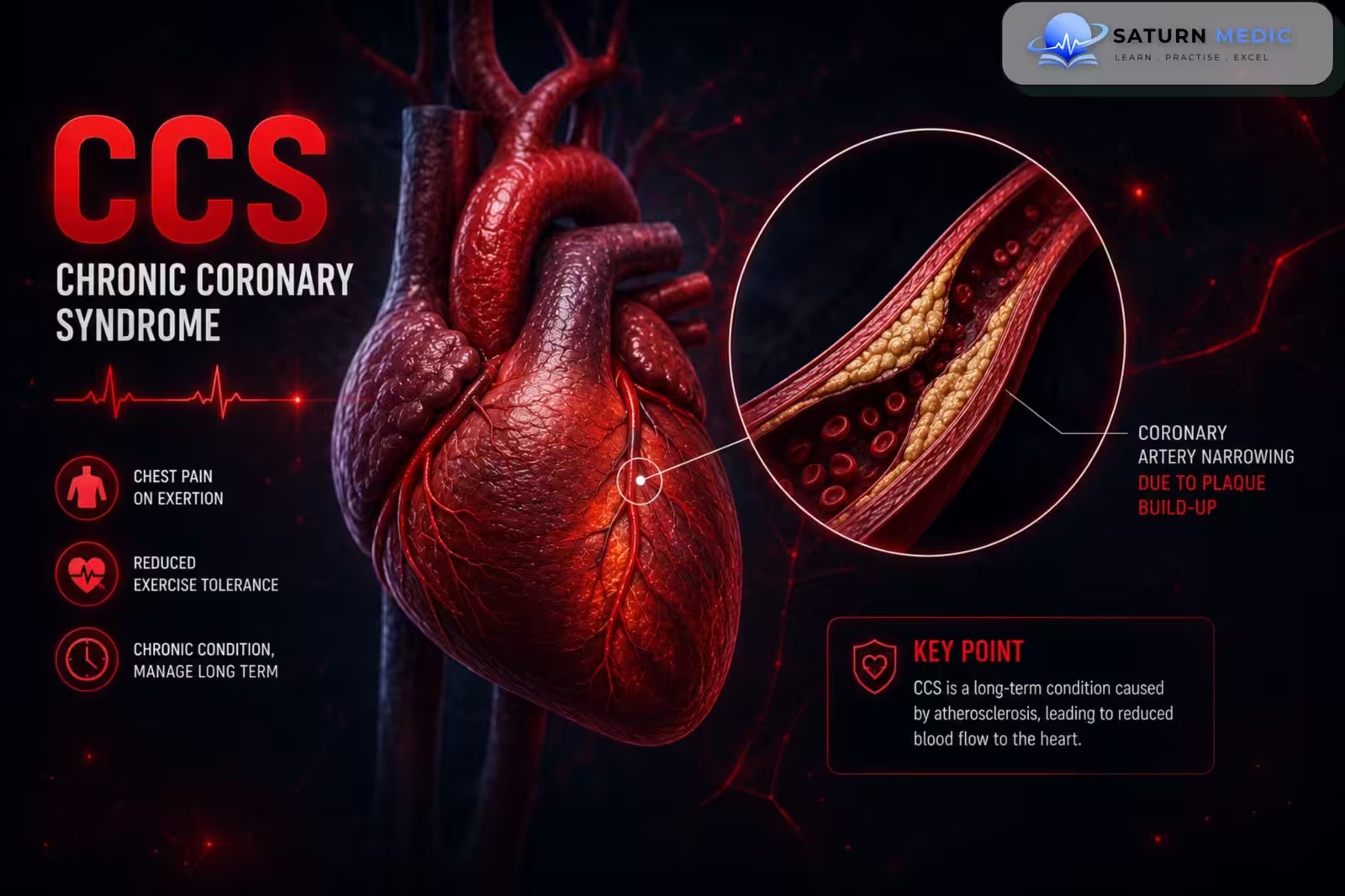
Chronic Coronary Syndrome (CCS) represents the stable, predictable phases of coronary artery disease, where a partial blockage restricts blood flow to the heart muscle during times of increased demand (like exercise)
Includes
• Stable Angina
• Microvascular or Vasospastic Angina
Etiology & Risk Factors
-The primary cause of Chronic Coronary Syndrome (CCS) is atherosclerosis (the gradual buildup of fatty plaques inside the coronary arteries). Over decades, cholesterol, inflammatory cells, and fibrous tissue form a stable plaque within the arterial wall. This narrows the lumen of the vessel.
Risk Factors ;
-Non Modifiable Risk Factors
These are the baseline traits that determine a patient’s genetic and physiological susceptibility:
Age: Risk increases progressively with age (commonly presenting in men > 45 and women > 55).
Male Sex: Men are generally at a higher risk, and women’s risk increases significantly post-menopause.
Family History: A strong history of premature cardiovascular disease (defined as a first-degree male relative affected before age 55, or a female relative before age 65).
Ethnicity: Certain populations, particularly individuals of South Asian descent, have a significantly higher baseline risk of premature coronary disease.
-Modifiable Risk Factors
These are the core focus of clinical prevention and treatment in any case study:
Dyslipidaemia: Elevated Low-Density Lipoprotein Cholesterol (LDL-C or “bad” cholesterol) and low High-Density Lipoprotein Cholesterol (HDL-C).
Hypertension: Chronic high blood pressure increases mechanical stress on arterial walls, accelerating plaque formation.
Cigarette Smoking: Directly damages the vascular endothelium, promotes plaque instability, and causes temporary vasoconstriction.
Diabetes Mellitus (and Insulin Resistance): Substantially accelerates atherosclerosis; diabetic patients frequently experience “silent” ischaemia due to autonomic neuropathy.
Obesity & Physical Inactivity: Contributes directly to metabolic syndrome, hypertension, and insulin resistance.
Pathophysiology
1. The Spark: Endothelial Injury & Inflammation
The process begins when the inner lining of the coronary artery (the endothelium) suffers chronic damage from systemic risk factors like smoking, hypertension, diabetes, or elevated LDL cholesterol. This structural breach allows Low-Density Lipoprotein (LDL) particles to penetrate deep into the arterial wall, where they become oxidized. In response, the immune system triggers an inflammatory cascade, deploying macrophages to engulf these lipids. These cells transform into lipid-laden foam cells, which aggregate to form the foundational “fatty streak” of the disease.
2. The Build-up: Fibrous Cap & Stable Plaque
Over years, smooth muscle cells migrate to the injury site and proliferate, secreting a dense matrix of collagen to form a firm, protective fibrous cap over the growing lipid core. This cap keeps the plaque physically “stable”—preventing it from suddenly rupturing and causing an emergency heart attack. However, as this stable plaque progressively expands inward, it creates a fixed, mechanical narrowing of the arterial lumen.
3. The Balance: Sufficiency at Rest
Under baseline conditions (when the patient is resting or calm), the cross-sectional area of the narrowed artery is still wide enough to allow adequate blood flow. The heart muscle receives exactly the amount of oxygen it needs for its basic metabolic demands, which is why the patient remains completely asymptomatic during normal, low-intensity daily life.
4. The Mismatch: Ischaemia and the Anginal Pain
The entire dynamic changes during physical exertion or acute emotional stress. The sympathetic nervous system activates, causing a surge in heart rate and myocardial contractility to pump more blood. This drives up the heart muscle’s demand for oxygen. Because the fixed atherosclerotic plaque physically prevents the coronary artery from dilating to supply more blood, a sudden supply-demand mismatch occurs.
Clinical Presentation
1. Symptoms: The Classic Triad of Stable Angina
The diagnosis of stable angina relies heavily on identifying the three hallmark characteristics of ischemic chest pain:
Location & Character: A sensation of pressure, tightness, squeezing, or heaviness in the center of the chest (retrosternal). It is rarely a sharp, stabbing, or localized pain.
Radiation: The discomfort commonly radiates to the neck, jaw, epigastrium (upper abdomen), or down one or both arms (typically the left ulnar border).
Precipitating Factors: The pain is predictably brought on by situations that increase myocardial oxygen demand, such as physical exertion (walking uphill or against a cold wind) or intense emotional stress.
Relieving Factors: The symptoms are transient, typically easing completely within 3 to 5 minutes of rest or rapidly after administering sublingual glyceryl trinitrate (GTN) spray/tablets.
Clinical Classification (The ESC/Davidson’s Criteria)
To classify the chest pain in your case study, use this standard diagnostic tool:
Typical Angina: Meets all 3 of the characteristics above.
Atypical Angina: Meets only 2 of the characteristics above.
Non-Anginal Chest Pain: Meets 1 or none of the characteristics.
2. Associated Symptoms
Some patients, particularly older individuals, women, or those with long-standing diabetes, may not present with classic chest pain. Instead, they present with anginal equivalents:
Dyspnoea (Shortness of Breath): Often the primary or only symptom of ischemia due to transiently increased left ventricular stiffness during exercise.
Autonomic Symptoms: Nausea, mild sweating (diaphoresis), or unexplained fatigue during exertion
History Taking
Diagnosis rests on the combination of clinical history, ECG findings, and cardiac biomarkers.
Key Questions
Character of chest pain
Onset and duration
Radiation
Presence at rest or with exertion
Previous episodes
Response to nitroglycerin
Associated dyspnea
Nausea or vomiting
Syncope
Palpitations
Red Flags
Hemodynamic instability
Acute pulmonary edema
Tachyarrhythmias
Bradyarrhythmia
Cardiogenic shock
Physical Examination
During a stable period, the physical examination is frequently unremarkable. However, looking for specific signs that point to underlying risk factors or structural complications:
Cardiovascular Signs
Hypertension: Elevated blood pressure readings.
Cardiac Murmurs: An ejection systolic murmur might suggest aortic stenosis (a key differential or aggravating factor), while a pansystolic murmur could indicate ischemic mitral regurgitation.
Signs of Heart Failure: Manifesting as a third heart sound (S3), raised Jugular Venous Pressure (JVP), or basal lung crackles, indicating severe underlying CAD.
Metabolic Signs (Hyperlipidaemia)
Xanthelasmata: Yellowish cholesterol deposits on the eyelids.
Tendon Xanthomas: Lipid deposits over the Achilles or extensor tendons.
Corneal Arcus: A white or grey ring around the cornea (significant if the patient is under 50 years old).
Vascular Signs
Peripheral Arterial Disease (PAD): Diminished peripheral pulses (e.g., dorsalis pedis) or carotid bruits, indicating widespread systemic atherosclerosis.
Long-term secondary prevention typically includes dual antiplatelet therapy, a high-intensity statin, a beta-blocker, an ACE inhibitor or ARB (especially with reduced ejection fraction), and structured cardiac rehabilitation.
Investigations
According to Davidson’s Principles and Practice of Medicine, investigations for suspected Chronic Coronary Syndrome (CCS) are structured to achieve three goals: confirm the diagnosis of ischemia, rule out alternative causes of chest pain, and assess the patient’s long-term cardiovascular risk.
1. First-Line Diagnostic Investigations (To Confirm CCS)
Modern guidelines heavily favor non-invasive anatomical imaging over traditional exercise stress tests for initial diagnosis:
CT Coronary Angiography (CTCA): This is now the preferred first-line investigation for patients with atypical or typical chest pain. It provides a highly accurate, non-invasive anatomical view of the coronary arteries to detect calcification and plaque blockages.
Non-Invasive Functional Imaging: If CTCA is unavailable, inconclusive, or shows blockages of uncertain clinical significance, functional imaging is used to look for actual myocardial ischemia during stress. These include:
Stress Echocardiography (using exercise or dobutamine).
Myocardial Perfusion Scintigraphy (MPS / SPECT).
Stress Cardiac MRI.
2. Baseline Laboratory Tests (To Assess Risk Factors)
These blood tests look for underlying causes, comorbidities, and metabolic risk profiles:
Fasting Lipid Profile: To measure total cholesterol, LDL-C, HDL-C, and triglycerides.
HbA1c / Fasting Plasma Glucose: To screen for or monitor Diabetes Mellitus.
Full Blood Count (FBC): To rule out anemia, which can precipitate or worsen angina by decreasing oxygen delivery.
Renal Function (U&Es / eGFR): To check for Chronic Kidney Disease, which accelerates CAD and influences medication dosing.
Thyroid Function Tests (TFTs): To rule out hyperthyroidism (which increases heart rate/cardiac workload) or hypothyroidism (which worsens dyslipidemia).
3. Standard Baseline Cardiac Tests
12-Lead Resting ECG: Often entirely normal between episodes of pain. However, it may show signs of past silent myocardial infarctions (pathological Q-waves), left ventricular hypertrophy (LVH), or baseline ST-segment/T-wave abnormalities.
Echocardiogram (at rest): Done to assess Left Ventricular (LV) ejection fraction, evaluate regional wall motion abnormalities (suggesting old ischemic damage), and rule out structural heart diseases like aortic stenosis.
4. Invasive Gold Standard (For High-Risk Patients)
Invasive Coronary Angiography (ICA): Reserved for patients identified as high risk by non-invasive tests, those with severe symptoms refractory to medical therapy, or those being considered for immediate revascularization (stenting or bypass surgery).
Diagnosis
Angina · Clinical Comparison
+ antiplatelet + statin
β-blockers avoided — may worsen spasm
Management of Chronic Coronary Syndrome (CCS)
1. Medical Management (The Two-Track Therapy)
Track A: Prognostic Therapy (To Prevent Myocardial Infarction & Death)
These medications must be considered for all patients with confirmed CCS, regardless of their symptom frequency:
Antiplatelet Therapy: Aspirin (75–100 mg daily) is the first-line choice to prevent plaque thrombosis. If a patient is completely aspirin-intolerant, Clopidogrel (75 mg daily) is used as the alternative.
Lipid-Lowering Therapy: High-intensity Statins (e.g., Atorvastatin 40–80 mg) are initiated for all patients to stabilize coronary plaques, lower LDL cholesterol, and improve endothelial function.
ACE Inhibitors (or ARBs): Strongly recommended if the patient has co-existing conditions like hypertension, diabetes, chronic kidney disease, or left ventricular dysfunction (ejection fraction less than 40%).
Track B: Anti-Anginal Therapy (To Relieve Symptoms)
These therapies aim to resolve ischemia by lowering myocardial oxygen demand or enhancing coronary blood supply:
Immediate Relief: Sublingual Glyceryl Trinitrate (GTN) spray or tablets are prescribed to abort acute anginal attacks as they occur or as a preventative measure immediately before predictable physical exertion.
First-Line Preventative Options: * Beta-blockers (beta blockers): (e.g., Bisoprolol, Metoprolol). These lower heart rate and contractility, lowering oxygen demand. They are the preferred first-line anti-anginal agents.
Calcium Channel Blockers (CCBs): Rate-limiting CCBs (e.g., Verapamil, Diltiazem) can be used as an alternative to beta-blockers. Alternatively, dihydropyridine CCBs (e.g., Amlodipine) can be combined safely with beta-blockers if symptoms remain uncontrolled.
Second-Line Preventative Options: Added if first-line drugs are contraindicated, poorly tolerated, or insufficient:
Long-acting Nitrates (e.g., Isosorbide Mononitrate).
Ivabradine (selectively lowers heart rate by acting on the sinus node).
Nicorandil or Ranolazine.
2. Lifestyle Modification & Risk Factor Control
aggressive lifestyle management underpins all medical interventions:
Smoking Cessation: Complete cessation significantly reduces cardiovascular mortality.
Dietary Adjustments: Adopting a Mediterranean-style diet high in whole grains, vegetables, and unsaturated fats, while strictly limiting saturated fats and sodium.
Physical Activity: 30–60 minutes of moderate-intensity aerobic exercise most days of the week, tailored carefully to the patient’s ischemic threshold.
Weight & Metabolic Control: Managing obesity and ensuring tight glycemic control in patients with diabetes (aiming for an individualized HbA1c target).
3. Revascularization Therapy
If a patient’s symptoms are poorly controlled despite optimal medical therapy, or if non-invasive investigations indicate a high risk of a major cardiac event, mechanical revascularization is indicated:
Percutaneous Coronary Intervention (PCI): Involves dilating the narrowed artery with a balloon and deploying a Drug-Eluting Stent (DES) to keep the vessel open. This is highly effective at relieving symptoms but is typically reserved for localized, less complex blockages.
Coronary Artery Bypass Grafting (CABG): A surgical procedure using arterial or venous grafts (such as the internal mammary artery or saphenous vein) to bypass severe blockages. It is favored for patients with complex multivessel disease, left main stem stenosis, or concurrent severe left ventricular dysfunction.
Complications of Chronic Coronary Syndrome (CCS)
-Acute Coronary Syndrome (ACS): Stable plaque ruptures or erodes, causing sudden thrombosis that leads to Unstable Angina, NSTEMI, or STEMI (Heart Attack).
-Heart Failure (HF): Chronic lack of oxygen causes muscle scarring (Ischaemic Cardiomyopathy) or forces muscle tissue to shut down to survive (Hibernating Myocardium), severely reducing pumping efficiency.
-Arrhythmias & Sudden Cardiac Death (SCD): Ischaemia disrupts electrical stability, triggering fatal rhythms like Ventricular Tachycardia (VT) or Ventricular Fibrillation (VF).
-Ischaemic Mitral Regurgitation: Ischaemia of the papillary muscles causes the mitral valve to leak, leading to acute pulmonary congestion.
Prognosis of Chronic Coronary Syndrome (CCS)
1. Chronic Coronary Syndrome (CCS)
Overall Outlook: Generally stable but carries a steady risk of progression. The annual mortality rate is 1% to 2%, and the annual risk of a non-fatal heart attack is about 3%.
Major Risks: The primary danger is a sudden plaque rupture leading to an Acute Coronary Syndrome (Heart Attack) or the gradual development of heart failure over time.
Survival Predictors: Survival is heavily determined by how many arteries are blocked, whether the Left Main artery is involved, and how well the left ventricle pumps blood (Ejection Fraction).
2. Vasospastic Angina (Prinzmetal’s)
Overall Outlook: Generally excellent in the short-to-medium term, carrying a significantly higher long-term survival rate than classic CCS (often $>90\%$ survival over 5 years) if treated properly.
Major Risks: Because there is usually no major underlying plaque block, the risk of a typical heart attack is much lower. Instead, the primary dangers are severe arrhythmias (like Ventricular Fibrillation) or sudden fainting triggered during an intense, prolonged spasm.
Survival Predictors: Survival depends almost entirely on avoiding triggers (like smoking or cocaine) and consistently taking Calcium Channel Blockers to prevent the arteries from spasming.
Key Points / Clinical Pearls of Chronic Coronary Syndrome (CCS)
- What it is: Chronic Coronary Syndrome (CCS) is a stable, predictable buildup of plaque in the heart’s arteries that narrows blood flow.
The Trigger: Chest pain (stable angina) happens only when the heart works harder (exercise, stress) and goes away with rest or medication.
Main Test: Doctors now prefer a CT Coronary Angiography (CTCA) scan to look inside the arteries, rather than the old treadmill walking test.
Treatment: Patients take medications to stop pain (Beta-blockers) and lifesaving drugs to prevent a heart attack (Aspirin and Statins).
Fixing the Blockage: Stents (PCI) are used to clear pain, but Bypass Surgery (CABG) is used to save lives in severe, multi-vessel blockages.
- National Center for Biotechnology Information (NIH). Acute Coronary Syndrome, StatPearls.
- Rao SV, O'Donoghue ML, Ruel M, et al. 2025 ACC/AHA/ACEP/NAEMSP/SCAI Guideline for the Management of Patients With Acute Coronary Syndromes. Circulation. 2025;151:e771-e862. doi: 10.1161/CIR.0000000000001309.
- MedlinePlus, National Library of Medicine (NIH). Acute Coronary Syndrome: Health Topic.
- National Center for Biotechnology Information (NIH). Selecting a Treatment Modality in Acute Coronary Syndrome, StatPearls.
- National Center for Biotechnology Information (NIH). Unstable Angina, StatPearls.