

Clinical Subject Page
Heart Failure with Reduced Ejection Fraction (HFrEF)
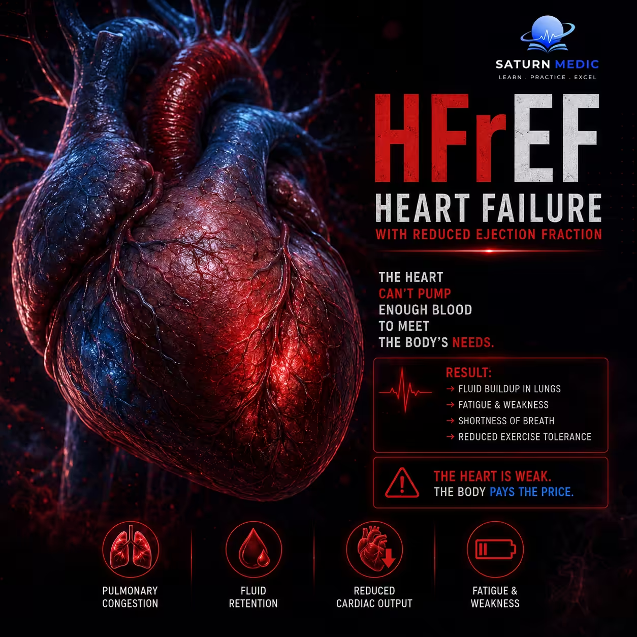
Commonly known as a Systolic Heart Failure — means the heart has a pumping problem, Because the pump is weak, blood pools inside the heart and backs up into the lungs and body
Also called
ICD-10
Specialty
Onset
Reviewed
-
OverviewOverview
-
Etiology & Risk FactorsEtiology & Risk Factors
-
PathophysiologyPathophysiology
-
Clinical PresentationClinical Presentation
-
History TakingHistory Taking
-
Physical ExaminationPhysical Examination
-
InvestigationsInvestigations
-
DiagnosisDiagnosis
-
ManagementManagement
-
ComplicationsComplications
-
PrognosisPrognosis
-
Key Points / Clinical PearlsKey Points / Clinical Pearls
Overview
Heart Failure with Reduced Ejection Fraction (HFrEF), historically called systolic heart failure, occurs when the left ventricle muscle becomes too weak or damaged to pump blood effectively. This leads to an Ejection Fraction (EF) of 40% or less, meaning the heart pumps out less than 40% of the blood filling its main chamber with each beat
Etiology & Risk Factors
Etiology (Underlying Causes)
Heart Failure with Reduced Ejection Fraction (HFrEF) occurs when a disease process directly damages or weakens the heart muscle (myocardium). The most common causes include:
Ischemic Heart Disease (Coronary Artery Disease): The single leading cause. A past myocardial infarction (heart attack) leaves behind non-functional scar tissue, permanently reducing the heart’s overall pumping power.
Dilated Cardiomyopathy (DCM): Genetic mutations, viral infections (myocarditis), or toxins cause the left ventricle to stretch out, thinned, and weaken.
Valvular Heart Disease: Long-standing aortic stenosis, aortic regurgitation, or mitral regurgitation forces the muscle to work too hard, eventually exhausting and dilating it.
Toxins & Medications: Chronic alcohol abuse, cocaine use, or exposure to cardiotoxic chemotherapy agents (e.g., anthracyclines like doxorubicin).
-Risk Factors ;
Hypertension (High Blood Pressure): Uncontrolled pressure forces the heart to pump against high resistance. While it initially causes thickening Heart Failure with Reduced Ejection Fraction (HFrEF), long-term stress can cause the muscle to fail and dilate.
Diabetes Mellitus: Directly damages micro-vessels in the heart and alters cardiac metabolism, multiplying the risk of heart failure.
Metabolic & Lifestyle Factors: Obesity, severe hyperlipidemia, smoking, and a sedentary lifestyle.
Sleep Apnea: Severe obstructive sleep apnea causes sudden shifts in intrathoracic pressure and oxygen drops, placing immense nightly stress on the left ventricle.
Advanced Age & Family History: A family history of cardiomyopathy or early-onset heart disease significantly elevates individual risk.
Pathophysiology
-
Initial Injury: A heart attack or chronic high blood pressure damages and weakens the heart muscle.
-
Systolic Dysfunction: The weakened left ventricle stretches out and loses its ability to pump forcefully (Ejection Fraction less than 40%).
-
Neurohormonal Activation: The body mistakes low blood flow for blood loss, triggering a massive release of adrenaline (SNS) and stress hormones (RAAS).
-
Vicious Cycle: These hormones force the weak heart to beat faster and clamp blood vessels tight to maintain blood pressure.
-
Cardiac Remodeling: Chronically high hormone levels backfire, causing the heart walls to permanently thin, stretch, and scar.
-
Backward Failure: The failing pump cannot clear blood, causing fluid to back up into the lungs (breathlessness) and legs (swelling).
-
Forward Failure: Decreased blood output starves the body’s vital organs of oxygen, causing severe fatigue and kidney dysfunction.
Clinical Presentation
1. Symptoms
Fluid Overload Symptoms (Backward Failure)
Dyspnoea (Shortness of Breath): Initially occurs only during exertion, but progressively worsens to occur at rest as fluid backs up into the lung alveoli (pulmonary congestion).
Orthopnoea: Shortness of breath that occurs when lying flat. Patients often report needing to sleep propped up on multiple pillows.
Paroxysmal Nocturnal Dyspnoea (PND): A highly specific sign where the patient wakes up suddenly at night gasping for air, usually 1–2 hours after falling asleep, due to the central redistribution of edema fluid.
Peripheral Edema: Bilateral, dependent swelling starting in the ankles and feet, which may progress up to the thighs, scrotum, and abdominal wall (anasarca).
Low Cardiac Output Symptoms (Forward Failure)
Fatigue and Exercise Intolerance: Due to poor skeletal muscle perfusion.
Dizziness, Lightheadedness, or Confusion: Occurs in advanced stages due to reduced cerebral blood flow.
Unexplained Weight Loss (Cardiac Cachexia): Wasting of muscle and tissue caused by a combination of low tissue perfusion, gut congestion (leading to poor nutrient absorption), and chronic inflammation.
–Clinical Classification : New York Heart Association Classification (NYHA) ;
Heart Failure · NYHA Functional Classification
| Class | Functional Limitation | Symptoms | 5-Year Mortality | ACC/AHA Stage |
|---|---|---|---|---|
|
I
No limitation
|
Ordinary physical activity causes no symptoms. No restriction on daily activities. | Asymptomatic despite cardiac disease | <10% | Stage C |
|
II
Slight limitation
|
Comfortable at rest. Ordinary activity (climbing stairs, walking briskly) causes fatigue, dyspnea, or palpitations. | Dyspnea on moderate exertion; orthopnea may be absent | ~20% | Stage C |
|
III
Marked limitation
|
Comfortable at rest. Less-than-ordinary activity (dressing, walking on flat) causes symptoms. | Dyspnea on minimal exertion, fatigue, orthopnea, possible PND | ~50% | Stage C |
|
IV
Symptoms at rest
|
Unable to carry on any activity without discomfort. Symptoms present at rest. Bed-to-chair existence. | Dyspnea at rest, severe orthopnea, PND, peripheral edema, ascites | >50% | Stage D |
EF-based Classification
History Taking
Key Questions for Heart Failure with Reduced Ejection Fraction (HFrEF)
-Fluid Congestion (Backward Failure)
Exertional Dyspnoea: Onset and severity during physical activity.
Orthopnoea: Presence of shortness of breath when lying flat, and the number of pillows required.
Paroxysmal Nocturnal Dyspnoea (PND): Sudden awakening at night gasping for air.
Peripheral Edema: New or worsening swelling in the ankles, legs, or abdomen.
Weight Changes: Rapid, unexpected weight gain over a few days (fluid accumulation).
-Low Cardiac Output (Forward Failure)
Fatigue: Severe, unusual exhaustion during basic daily tasks.
Cerebral Hypoperfusion: Dizziness, lightheadedness, or confusion, especially when standing.
-Red Flags for Heart Failure with Reduced Ejection Fraction (HFrEF)
• Hemodynamic instability
• Acute pulmonary edema
• Tachyarrhythmias
• Bradyarrhythmia
• Cardiogenic shock
Physical Examination
-Signs of Volume Overload (Fluid Retention)
Elevated Jugular Venous Pressure (JVP): Reflects high pressure in the right atrium. A positive hepatojugular reflux (pressing the liver raises JVP) is a highly reliable sign.
Pulmonary Crackles (Crepitations): Fine, inspiratory Velcro-like crackles heard at the lung bases, signaling fluid in the alveoli.
Pitting Peripheral Edema: Symmetrical swelling in the lower extremities that leaves an indentation when pressed.
-Signs of Cardiac Dysfunction (The Failing Pump)
Displaced Apex Beat: The apex beat is shifted downwards and outwards (lateral to the midclavicular line), indicating left ventricular dilation.
Third Heart Sound (S3 Gallop): A low-pitched extra heart sound heard early in diastole, caused by rapid blood flow rushing into a stiff, dilated, and volume-overloaded ventricle.
Tachycardia: A rapid heart rate at rest as the sympathetic nervous system tries to compensate for the low stroke volume.
Investigations
1. Core Diagnostic Tests
Transthoracic Echocardiogram (Echo): The most important test. It uses ultrasound to view the heart and measure the Left Ventricular Ejection Fraction (LVEF). A value of less than 40% confirms HFrEF. It also checks for valve problems and chamber enlargement.
Natriuretic Peptides (BNP or NT-proBNP): A blood test used to rule out heart failure. If a patient is short of breath but their BNP is normal, heart failure is highly unlikely. High levels mean the heart muscle is stretched and under stress.
12-Lead Electrocardiogram (ECG): Used to find the underlying cause of heart failure. It looks for signs of a past heart attack (Q waves), a thickened heart wall, or irregular rhythms like Atrial Fibrillation.
Chest X-ray (CXR): Checks the lungs for fluid buildup. It can show an enlarged heart silhouette (cardiomegaly) and fluid in or around the lungs (pulmonary edema).
2. Routine Blood Tests
Kidney Function & Electrolytes (U&Es): Checks creatinine and potassium levels. This is vital because standard heart failure medications directly affect the kidneys and alter potassium levels.
Full Blood Count (FBC): Screens for anemia, which forces a weak heart to work harder and worsens symptoms.
Thyroid Function Tests (TFTs): Rules out thyroid issues, as an overactive or underactive thyroid can trigger or worsen heart failure.
3. Advanced Testing
Cardiac MRI (CMR): Used if the Echo images are unclear or to find the exact cause of the muscle weakness (e.g., distinguishing between a past silent heart attack and inflammation).
Coronary Angiography: Performed if the patient has chest pain or if blocked arteries are suspected to be the root cause of the heart failure.
Diagnosis
Diagnosis · HFrEF vs HFpEF
EF < 40%
EF ≥ 50%
EF 40–49% = HFmrEF (grey zone)
• Reduced wall motion (global hypokinesia)
• Thin LV walls (eccentric remodelling)
• MR ± TR (secondary)
• LVH — concentric remodelling
• Impaired relaxation: E/e′ >14, e′ <7 cm/s
• LA dilation (LAVi >34 mL/m²)
BNP >100 pg/mL
NT-proBNP >300 pg/mL
BNP >35 pg/mL
NT-proBNP >125 pg/mL
LV dilation pattern; AF common
May be normal; ST changes of LVH strain
• High LVEDP
• High PCWP (>18 mmHg)
• High LVEDP (diastolic)
• PCWP rises steeply on exertion
ICD/CRT if indicated
Diuretics for congestion
No proven mortality benefit for RAAS/BB
Management of Heart Failure with Reduced Ejection Fraction (HFrEF)
1. The “Fantastic Four” (Saves Lives)
These four medications target the body’s destructive neurohormonal pathways. They should be started early and increased slowly to the maximum tolerated dose.
ARNI (Sacubitril-Valsartan): The preferred first-line drug. It relaxes blood vessels and helps the body flush fluid. Note: Never take within 36 hours of an ACE inhibitor.
Beta-blockers (e.g., Bisoprolol): Protects the heart from chronic adrenaline stress. It lowers the heart rate so the heart can pump more efficiently. Note: Only start when the patient is stable and fluid-free.
MRAs (e.g., Spironolactone): Blocks aldosterone to stop tissue scarring and fluid retention. Note: Requires regular blood tests to check potassium levels.
SGLT2 Inhibitors (e.g., Dapagliflozin): Originally for diabetes, this drug helps the kidneys excrete salt and improves heart energy, heavily reducing hospitalization risk.
2. Diuretics (Relieves Symptoms)
Loop Diuretics (e.g., Furosemide): The core treatment for symptom relief. They force the kidneys to dump excess salt and water, clearing fluid from the lungs and legs.
Note: Diuretics make patients feel better but do not improve long-term survival. Doses are adjusted based on daily weight.
3. Devices (For Advanced Stages)
Considered if the ejection fraction remains less than 35% after 3 months of optimal drug therapy.
ICD (Defibrillator): Monitors the heart and delivers an electrical shock if a lethal, rapid heart rhythm occurs, preventing sudden cardiac death.
CRT (Biventricular Pacemaker): Resynchronizes the left and right ventricles so they pump together in a coordinated, powerful squeeze.
Complications of Heart Failure with Reduced Ejection Fraction (HFrEF)
Acute Decompensated Heart Failure (ADHF): Sudden, severe worsening of symptoms (severe breathlessness, hypoxia) requiring urgent hospitalization and intravenous diuretics.
Arrhythmias: Damaged heart tissue disrupts the heart’s electrical system. Atrial Fibrillation is highly common and worsens pump failure, while Ventricular Tachycardia/Fibrillation can cause sudden cardiac death.
Renal Failure (Cardiorenal Syndrome): Poor forward blood flow from the heart starves the kidneys of oxygen, while backward fluid congestion increases pressure in renal veins, causing progressive kidney damage.
Thromboembolism: Sluggish blood flow within the dilated, poorly contracting left ventricle increases the risk of blood clots forming, which can travel to the brain and cause a stroke.
Cardiac Cachexia: Severe, involuntary muscle and tissue wasting caused by high systemic inflammation, increased metabolic demands, and poor nutrient absorption due to a congested gut.
Prognosis of Heart Failure with Reduced Ejection Fraction (HFrEF)
The 5-Year Survival Trap: Despite advances in treatment, HFrEF carries a poor prognosis. Roughly 50% of patients die within 5 years of their initial diagnosis, making its survival rate worse than many common cancers.
-Predictors of Poor Prognosis:
Higher NYHA Class (Class III or IV).
- Advanced age.
Recurrent hospitalizations for acute failure.
Co-existing conditions like chronic kidney disease or diabetes.
Markedly low Ejection Fraction ($\le 25\%$) or persistently high BNP/NT-proBNP blood levels.
Key Points / Clinical Pearls
Heart Failure with Reduced Ejection Fraction (HFrEF) : The heart muscle is too weak to pump enough blood, diagnosed by an Ejection Fraction less 40%
Top Cause: A past heart attack that left behind damaged, scarred heart muscle.
Key Symptoms: Shortness of breath (especially when lying flat), swollen ankles, and severe fatigue.
Red Flags: Shortness of breath while completely at rest, sudden fainting, or chest pain.
Diagnosis: Confirmed using a heart ultrasound (Echocardiogram) and a blood test (BNP).
Treatment: The “Fantastic Four” medications save lives, while water pills (Furosemide) clear fluid.
Outlook: It is highly dangerous, with a 50% mortality rate within 5 years of diagnosis.
- Golla MSG, Brown KN, Gupta N. National Center for Biotechnology Information (NIH). Heart Failure and Ejection Fraction, StatPearls.
- Heidenreich PA, Bozkurt B, Aguilar D, et al. 2022 AHA/ACC/HFSA Guideline for the Management of Heart Failure. Circulation. 2022;145:e895-e1032. PMID: 35363499.
- Bhagat AM, Rezaie SR, Chen A, Bhalla V. Guideline-Directed Medical Therapy for the Treatment of Heart Failure With Reduced Ejection Fraction. Curr Opin Cardiol. 2023. PMID: 37254024.
- Optimization and Real-World Implementation of Guideline-Directed Medical Therapy in Heart Failure With Reduced Ejection Fraction: A Contemporary Clinical Review. PMC13094103.
- MedlinePlus, National Library of Medicine (NIH). Heart Failure: Health Topic.