

Clinical Subject Page
Heart Failure with Preserved Ejection Fraction (HFpEF)
Commonly known as a Diastolic Heart Failure — filling problem rather than a pumping problem
Also called
ICD-10
Specialty
Onset
Reviewed
-
OverviewOverview
-
Etiology & Risk FactorsEtiology & Risk Factors
-
PathophysiologyPathophysiology
-
Clinical PresentationClinical Presentation
-
History TakingHistory Taking
-
Physical ExaminationPhysical Examination
-
InvestigationsInvestigations
-
DiagnosisDiagnosis
-
ManagementManagement
-
ComplicationsComplications
-
PrognosisPrognosis
-
Key Points / Clinical PearlsKey Points / Clinical Pearls
Overview
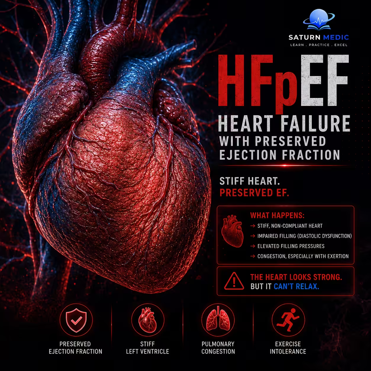
Heart Failure with Preserved Ejection Fraction (HFpEF) is a heart failure type where the heart muscle is too thick and stiff to relax and fill with enough blood, even though it can still squeeze and pump blood out normally.
Etiology & Risk Factors
Heart Failure with Preserved Ejection Fraction (HFpEF) usually results from impaired left ventricular relaxation and increased ventricular stiffness, leading to elevated filling pressures despite a relatively preserved left ventricular ejection fraction.
Common underlying causes include:
- Long-standing hypertension → left ventricular hypertrophy and increased stiffness
- Coronary artery disease / myocardial ischemia
- Atrial fibrillation
- Hypertrophic or infiltrative cardiomyopathies
- Valvular heart disease, especially aortic stenosis
- Diabetes mellitus
- Obesity
- Chronic kidney disease CKD
- Less commonly, restrictive pericardial or myocardial disease
-Risk Factors
- Older age
- Long-standing hypertension
- Obesity
- Diabetes mellitus
- Atrial fibrillation
- Coronary artery disease
- Chronic kidney disease CKD
- Female sex
Pathophysiology
Long-standing hypertension/obesity/diabetes or other cardiac disease → left ventricular hypertrophy and myocardial fibrosis → increased ventricular stiffness + impaired relaxation → reduced ventricular compliance → increased left ventricular filling pressure → increased left atrial pressure → pulmonary venous congestion → dyspnea and exercise intolerance → systemic venous congestion → peripheral edema
Simple Exam Sequence
Risk factors → LV hypertrophy/fibrosis → impaired relaxation + increased stiffness → ↑ filling pressure → pulmonary congestion → Heart Failure with Preserved Ejection Fraction (HFpEF)
Clinical Presentation
The Key Symptoms (What the Patient Feels)
Exertional Dyspnea (Shortness of Breath): This is the hallmark. Patients feel fine sitting still, but walking to the mailbox feels like climbing Mount Everest. Because the stiff heart cannot stretch to accommodate increased blood flow during exercise, pressure spikes instantly and floods the lungs.
Orthopnea & PND: The patient cannot sleep flat without suffocating (orthopnea) and often wakes up gasping for air a few hours after falling asleep (Paroxysmal Nocturnal Dyspnea). When lying down, fluid from the legs redistributes to the chest, overloading the stiff heart.
Profound Fatigue: Because the heart cannot rev its engine or increase its stroke volume, muscles are chronically starved of oxygenated blood.
Heart Failure · NYHA Functional Classification
| Class | Functional Limitation | Symptoms | 5-Year Mortality | ACC/AHA Stage |
|---|---|---|---|---|
|
I
No limitation
|
Ordinary physical activity causes no symptoms. No restriction on daily activities. | Asymptomatic despite cardiac disease | <10% | Stage C |
|
II
Slight limitation
|
Comfortable at rest. Ordinary activity (climbing stairs, walking briskly) causes fatigue, dyspnea, or palpitations. | Dyspnea on moderate exertion; orthopnea may be absent | ~20% | Stage C |
|
III
Marked limitation
|
Comfortable at rest. Less-than-ordinary activity (dressing, walking on flat) causes symptoms. | Dyspnea on minimal exertion, fatigue, orthopnea, possible PND | ~50% | Stage C |
|
IV
Symptoms at rest
|
Unable to carry on any activity without discomfort. Symptoms present at rest. Bed-to-chair existence. | Dyspnea at rest, severe orthopnea, PND, peripheral edema, ascites | >50% | Stage D |
EF-based Classification
History Taking
Key Questions
“How many pillows do you need to sleep on at night to breathe comfortably?”
“Do you ever wake up suddenly in the middle of the night gasping for air?”
“Have your shoes, socks, or pants been feeling tighter due to swelling in your legs or belly?”
“What is a daily activity you used to do easily that completely wipes you out now?”
“Do you have to stop and catch your breath just walking to the mailbox or going up one flight of stairs?”
“Are you experiencing a constant, crushing fatigue that doesn’t go away with rest?”
“Do you have a history of high blood pressure, diabetes, or kidney disease?”
“Have you ever been told you have an irregular or fluttering heartbeat (AFib)?”
-Red Flags for Heart Failure with Preserved Ejection Fraction (HFpEF)
• Hemodynamic instability
• Acute pulmonary edema
• Tachyarrhythmias
• Bradyarrhythmia
• Cardiogenic shock
Physical Examination
-The Physical Signs (What You See on Examination)
Jugular Venous Distension (JVD): The neck veins look bulging and distended. This is a direct visual map of the high pressure backing up from the right side of the heart.
Pulmonary Crackles (Rales): Listening to the lungs with a stethoscope reveals a crisp, crackling sound at the bases—the literal sound of air bubbling through fluid in the alveoli.
Pitting Peripheral Edema: Significant fluid retention causes swollen ankles and legs. Pressing a thumb into the shin leaves a persistent indentation.
-The Clinical Phenotype (The Typical Patient Profile)
You can often spot a potential HFpEF patient before they even open their mouth. Unlike HFrEF patients (who are often younger men with a history of a massive heart attack), the classic HFpEF patient is typically:
An older female (often 70+).
Living with long-standing, poorly controlled hypertension.
Managing metabolic syndrome (obesity and Type 2 diabetes).
Frequently in Atrial Fibrillation (AFib)—because the high pressure stretches the left atrium until its electrical pathways become chaotic.
Investigations
1. Biomarker Assessment (Natriuretic Peptides)
Natriuretic peptides are the first laboratory tests used when HFpEF is suspected. Increased myocardial wall stress caused by elevated cardiac filling pressures stimulates the release of B-type natriuretic peptide (BNP) and N-terminal pro-B-type natriuretic peptide (NT-proBNP). Elevated levels support the diagnosis of heart failure, whereas normal levels make HFpEF less likely. Because atrial fibrillation independently increases natriuretic peptide concentrations, higher diagnostic thresholds are required in patients with this arrhythmia.
2. Transthoracic Echocardiography
Transthoracic echocardiography is the most important non-invasive imaging test for diagnosing HFpEF. It confirms that the LVEF is preserved (≥50%) and evaluates both cardiac structure and diastolic function.
Key echocardiographic findings include:
Diastolic dysfunction: The E/e′ ratio estimates left ventricular filling pressure. A higher ratio indicates impaired ventricular relaxation and elevated filling pressures.
Left atrial enlargement: The left atrial volume index (LAVI) reflects chronic exposure to elevated filling pressures. Enlargement of the left atrium suggests long-standing diastolic dysfunction.
Left ventricular hypertrophy: The left ventricular mass index (LVMI) is used to detect increased ventricular wall thickness, which commonly results from chronic hypertension and contributes to ventricular stiffness.
3. Provocative Hemodynamic Testing
Some patients with HFpEF have normal filling pressures at rest but develop abnormal pressure increases during physical activity. When resting investigations are inconclusive but clinical suspicion remains high, additional testing is recommended.
Diastolic stress echocardiography: Echocardiographic measurements are obtained during exercise to identify exercise-induced elevations in left ventricular filling pressures that may not be present at rest.
Right heart catheterization: This is the gold standard for confirming HFpEF. It directly measures intracardiac and pulmonary capillary wedge pressures at rest or during exercise, providing definitive evidence of elevated left ventricular filling pressures when non-invasive tests are inconclusive.
Diagnosis
Diagnosis · HFrEF vs HFpEF
EF < 40%
EF ≥ 50%
EF 40–49% = HFmrEF (grey zone)
• Reduced wall motion (global hypokinesia)
• Thin LV walls (eccentric remodelling)
• MR ± TR (secondary)
• LVH — concentric remodelling
• Impaired relaxation: E/e′ >14, e′ <7 cm/s
• LA dilation (LAVi >34 mL/m²)
BNP >100 pg/mL
NT-proBNP >300 pg/mL
BNP >35 pg/mL
NT-proBNP >125 pg/mL
LV dilation pattern; AF common
May be normal; ST changes of LVH strain
• High LVEDP
• High PCWP (>18 mmHg)
• High LVEDP (diastolic)
• PCWP rises steeply on exertion
ICD/CRT if indicated
Diuretics for congestion
No proven mortality benefit for RAAS/BB
Related Topics
Management of Heart Failure with Preserved Ejection Fraction (HFpEF)
1. The First-Line Breakthrough: SGLT2 Inhibitors
These are the undisputed cornerstone of modern HFpEF management. Originally designed for diabetes, drugs like Empagliflozin or Dapagliflozin are now prescribed to all HFpEF patients, regardless of whether they have diabetes.
How they work: They force the kidneys to dump excess sugar and sodium through the urine. This reduces blood volume, eases the pressure on the heart, and drastically lowers the risk of hospitalization.
2. Fluid and Pressure Control (Symptom Relief)
Loop Diuretics (Furosemide / Bumetanide): These are the “water pills.” While they don’t fix the stiff heart itself, they are critical for pulling excess fluid out of the lungs and swollen legs to relieve breathlessness.
Targeted Blood Pressure Control: Keeping blood pressure strictly controlled (typically <130/80 {mmHg} is mandatory. Lowering the pressure in the arteries gives the stiff heart less resistance to push against.
3. Second-Line Options (For Selected Patients)
If a patient is still struggling despite an SGLT2 inhibitor, guidelines allow for a few specific add-on therapies:
MRA (Spironolactone): A mild, potassium-sparing water pill that helps reduce the formation of rigid scar tissue (fibrosis) in the heart muscle.
ARNI (Sacubitril/Valsartan): A specialized blood pressure medication that helps dilate blood vessels and reduce heart strain, proven particularly beneficial in female HFpEF patients.
4. Aggressive Comorbidity Management
Because HFpEF is a systemic metabolic disease, you cannot fix the heart without fixing the rest of the body:
Obesity (GLP-1 Receptor Agonists): Medications like Semaglutide have shown massive success in HFpEF patients with obesity, rapidly reducing heart inflammation, improving exercise capacity, and shedding excess weight.
Atrial Fibrillation (AFib): If the patient lapses into AFib, the heart loses its coordinated rhythm, which a stiff ventricle cannot handle. Aggressive rhythm control (medications or ablation) is prioritized.
Complications of Heart Failure with Preserved Ejection Fraction (HFpEF)
- Acute decompensated heart failure
- Pulmonary edema
- Atrial fibrillation and other atrial arrhythmias
- Pulmonary hypertension
- Right-sided heart failure
- Renal dysfunction
- Hepatic congestion
- Recurrent hospital admissions
- Reduced exercise capacity
- Frailty and reduced quality of life
- Thromboembolism, particularly with atrial fibrillation
- Cardiogenic shock — uncommon but possible in severe decompensation
Prognosis of Heart Failure with Preserved Ejection Fraction (HFpEF)
The Survival Rate: Roughly 50% of patients die within 5 years of diagnosis, making it just as dangerous as a weak-pump heart failure.
The Causes: Half of the deaths are from the heart failing completely or a stroke, while the other half are caused by the patient’s other conditions, like kidney failure or diabetes.
The Bad Signs: The outlook gets much worse if the patient develops an irregular heartbeat (AFib), kidney damage, or requires repeated hospital visits.
The Good News: Modern lifestyle medications (like SGLT2 inhibitors and weight-loss drugs) are actively changing these statistics, keeping patients out of the hospital and living longer.
Key Points / Clinical Pearls of Heart Failure with Preserved Ejection Fraction (HFpEF)
- Heart Failure with Preserved Ejection Fraction (HFpEF) stands for Heart Failure with Preserved Ejection Fraction. The heart squeezes normally, but it is too stiff to relax and fill up with enough blood.
- This causes severe shortness of breath, especially when walking, moving, or lying flat in bed, along with extreme fatigue and fluid swelling in the ankles, legs, and belly.
- To diagnose it, an echocardiogram must show the heart pumping out at least 50% of its blood while operating under high stiffness. A BNP or NT-proBNP blood test is also used to detect high stress levels in the heart muscle.
- SGLT2 inhibitor medications are the primary treatment used to protect the heart and reduce hospital visits. Water pills are used alongside them to flush out excess fluid and keep the lungs dry.
- Left untreated, the disease carries a tough 50% five-year survival rate, making early detection and treatment critical.
- Golla MSG, Brown KN, Gupta N. National Center for Biotechnology Information (NIH). Heart Failure and Ejection Fraction, StatPearls.
- Heidenreich PA, Bozkurt B, Aguilar D, et al. 2022 AHA/ACC/HFSA Guideline for the Management of Heart Failure. Circulation. 2022;145:e895-e1032. PMID: 35363499.
- Bhagat AM, Rezaie SR, Chen A, Bhalla V. Guideline-Directed Medical Therapy for the Treatment of Heart Failure With Reduced Ejection Fraction. Curr Opin Cardiol. 2023. PMID: 37254024.
- Optimization and Real-World Implementation of Guideline-Directed Medical Therapy in Heart Failure With Reduced Ejection Fraction: A Contemporary Clinical Review. PMC13094103.
- MedlinePlus, National Library of Medicine (NIH). Heart Failure: Health Topic.