

Clinical Subject Page
Acute Heart Failure (AHF)
Acute heart failure (AHF) is the rapid onset or sudden worsening of heart failure symptoms. It is a common medical emergency and often requires hospitalization.
Also called
ICD-10
Specialty
Onset
Reviewed
-
OverviewOverview
-
Etiology & Risk FactorsEtiology & Risk Factors
-
PathophysiologyPathophysiology
-
Clinical PresentationClinical Presentation
-
History TakingHistory Taking
-
Physical ExaminationPhysical Examination
-
InvestigationsInvestigations
-
DiagnosisDiagnosis
-
ManagementManagement
-
ComplicationsComplications
-
PrognosisPrognosis
-
Key Points / Clinical PearlsKey Points / Clinical Pearls
Overview
Acute Heart Failure (AHF) is the sudden onset or rapid worsening of heart failure symptoms due to the heart’s inability to pump enough blood to meet the body’s needs. It is a medical emergency that often requires hospitalization.
Etiology & Risk Factors
Etiology (Common Causes) for Acute Heart Failure (AHF)
Acute myocardial infarction (heart attack)
Acute decompensation of chronic heart failure
Uncontrolled hypertension
Cardiac arrhythmias (e.g., atrial fibrillation)
Valvular heart disease
Infection (e.g., pneumonia, sepsis)
Pulmonary embolism
Myocarditis
Renal failure or fluid overload
Medication or dietary nonadherence (e.g., excess sodium/fluid intake)
-Risk Factors ;
Pre-existing heart failure
Coronary artery disease
Hypertension
Diabetes mellitus
Chronic kidney disease
Advanced age
Obesity
Smoking
Excessive alcohol or illicit drug use (e.g., cocaine)
History of myocardial infarction or cardiomyopathy
Valvular heart disease
Pathophysiology
Acute cardiac dysfunction or sudden worsening of chronic heart failure → reduced cardiac output and/or increased ventricular filling pressures → activation of sympathetic nervous system and renin–angiotensin–aldosterone system → vasoconstriction + sodium and water retention → increased preload and afterload → further rise in cardiac filling pressures → pulmonary and/or systemic congestion → acute heart failure symptoms
Simple Exam Sequence
Trigger → acute rise in filling pressures ± reduced cardiac output → pulmonary/systemic congestion → dyspnea, edema, hypoperfusion
Clinical Presentation
Symptoms
Acute dyspnea
Orthopnea
Paroxysmal nocturnal dyspnea
Cough, sometimes with frothy sputum
Fatigue and weakness
Reduced exercise tolerance
Chest pain when ischemia is the precipitant
Palpitations
Reduced urine output
Rapid weight gain from fluid retention
Signs
Tachypnea
Tachycardia
Hypoxemia
Elevated JVP
Pulmonary crackles
Peripheral edema
S3 gallop
Cool extremities in low-output states
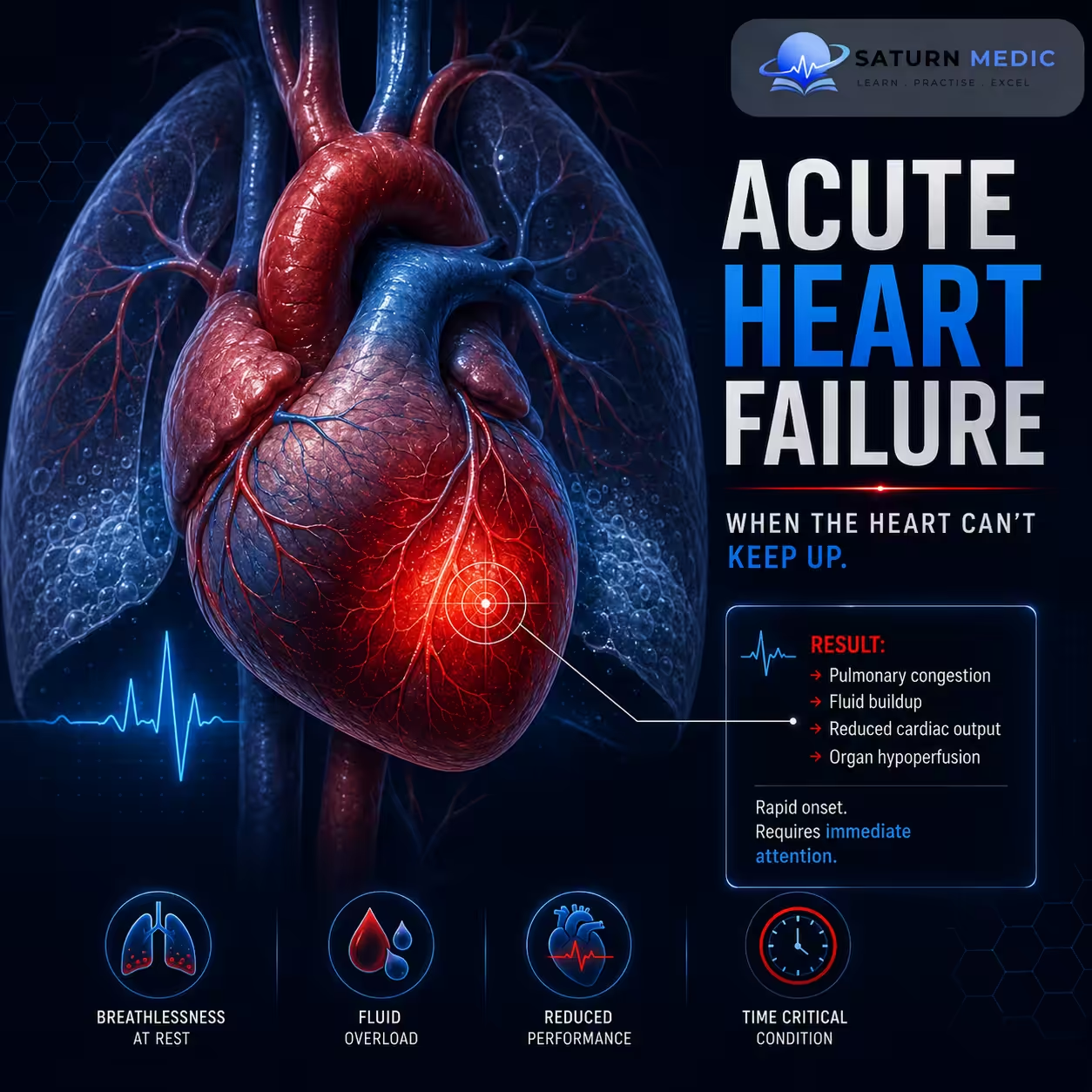
History Taking
Key Questions
-Fluid Congestion (Backward Failure)
Exertional Dyspnoea: Onset and severity during physical activity.
Orthopnoea: Presence of shortness of breath when lying flat, and the number of pillows required.
Paroxysmal Nocturnal Dyspnoea (PND): Sudden awakening at night gasping for air.
Peripheral Edema: New or worsening swelling in the ankles, legs, or abdomen.
Weight Changes: Rapid, unexpected weight gain over a few days (fluid accumulation).
-Low Cardiac Output (Forward Failure)
Fatigue: Severe, unusual exhaustion during basic daily tasks.
Cerebral Hypoperfusion: Dizziness, lightheadedness, or confusion, especially when standing.
-Red Flags
• Hemodynamic instability
• Acute pulmonary edema
• Tachyarrhythmias
• Bradyarrhythmia
• Cardiogenic shock
Physical Examination
-Signs of Volume Overload (Fluid Retention)
Elevated Jugular Venous Pressure (JVP): Reflects high pressure in the right atrium. A positive hepatojugular reflux (pressing the liver raises JVP) is a highly reliable sign.
Pulmonary Crackles (Crepitations): Fine, inspiratory Velcro-like crackles heard at the lung bases, signaling fluid in the alveoli.
Pitting Peripheral Edema: Symmetrical swelling in the lower extremities that leaves an indentation when pressed.
-Signs of Cardiac Dysfunction (The Failing Pump)
Displaced Apex Beat: The apex beat is shifted downwards and outwards (lateral to the midclavicular line), indicating left ventricular dilation.
Third Heart Sound (S3 Gallop): A low-pitched extra heart sound heard early in diastole, caused by rapid blood flow rushing into a stiff, dilated, and volume-overloaded ventricle.
Tachycardia: A rapid heart rate at rest as the sympathetic nervous system tries to compensate for the low stroke volume.
Investigations
- BNP or NT-proBNP – Elevated, supports the diagnosis of heart failure.
- Cardiac troponin – Detects myocardial injury or acute myocardial infarction.
- ECG (Electrocardiogram) – Identifies arrhythmias, ischemia, or infarction.
- Chest X-ray – Shows pulmonary edema, cardiomegaly, or pleural effusion.
- Echocardiogram – Assesses cardiac structure, ejection fraction, and valvular function.
- Blood tests: CBC, electrolytes, renal function (BUN/creatinine), liver function, glucose, and thyroid function (when indicated).
- Arterial blood gas (ABG) – Evaluates oxygenation and acid–base status in severe respiratory distress.
- Pulse oximetry – Monitors oxygen saturation continuously.
Diagnosis
Acute Heart Failure · Diagnostic Criteria — ESC 2021
| Clinical Profile | Congestion | Perfusion | Haemodynamics | Management focus |
|---|---|---|---|---|
| Warm & Dry | No congestion | Adequate perfusion | Normal CO, normal PCWP | Optimise oral therapy; not acute HF presentation |
| Warm & Wet | Congested | Adequate perfusion | Normal CO, high PCWP | Most common AHF. IV diuretics (furosemide), vasodilators, O₂ |
| Cold & Wet | Congested | Hypoperfused | Low CO, high PCWP | Inotropes + diuretics; consider haemodynamic monitoring; ICU |
| Cold & Dry | No congestion | Hypoperfused | Low CO, low/normal PCWP | Cardiogenic shock / RV failure. Cautious fluids + inotropes; urgent echo |
Heart Failure With Reduced Ejection Fraction (HFrEF) Vs Heart Failure With Preserved Ejection Fraction (HFpEF)
Diagnosis · HFrEF vs HFpEF
EF < 40%
EF ≥ 50%
EF 40–49% = HFmrEF (grey zone)
• Reduced wall motion (global hypokinesia)
• Thin LV walls (eccentric remodelling)
• MR ± TR (secondary)
• LVH — concentric remodelling
• Impaired relaxation: E/e′ >14, e′ <7 cm/s
• LA dilation (LAVi >34 mL/m²)
BNP >100 pg/mL
NT-proBNP >300 pg/mL
BNP >35 pg/mL
NT-proBNP >125 pg/mL
LV dilation pattern; AF common
May be normal; ST changes of LVH strain
• High LVEDP
• High PCWP (>18 mmHg)
• High LVEDP (diastolic)
• PCWP rises steeply on exertion
ICD/CRT if indicated
Diuretics for congestion
No proven mortality benefit for RAAS/BB
Management of Acute Heart Failure (AHF)
Immediate Management of Acute Heart Failure (AHF)
Airway, Breathing, Circulation (ABCs)
Supplemental oxygen if hypoxemic
Non-invasive ventilation (CPAP/BiPAP) for severe respiratory distress or pulmonary edema
Continuous cardiac and pulse oximetry monitoring
IV access and close monitoring of vital signs
Pharmacologic Treatment
IV loop diuretics (e.g., furosemide) for fluid overload
IV vasodilators (e.g., nitroglycerin) if blood pressure is adequate and congestion is present
Inotropes/vasopressors (e.g., dobutamine, norepinephrine) for cardiogenic shock or severe hypoperfusion
Treat the underlying cause (e.g., acute myocardial infarction, arrhythmia, infection, hypertensive emergency)
Ongoing Management for Acute Heart Failure (AHF)
Monitor urine output, daily weight, renal function, and electrolytes
Restrict sodium and fluids if indicated
Optimize guideline-directed heart failure medications once the patient is hemodynamically stable
Provide patient education and arrange appropriate follow-up to reduce the risk of readmission.
Complications of Acute Heart Failure (AHF)
- Acute pulmonary edema
- Cardiogenic shock
- Acute respiratory failure
- Hypoxemia
- Acute Kidney Injury (AKI)
- Worsening renal dysfunction
- Hepatic congestion or ischemic hepatitis
- Cardiac arrhythmias
- Myocardial ischemia or infarction
- Thromboembolism
- Severe electrolyte disturbances
- Metabolic acidosis
- Multiorgan failure
Prognosis of Acute Heart Failure (AHF)
-
- Prognosis depends on the underlying cause, severity, age, comorbidities, and response to treatment.
- Early diagnosis and prompt treatment improve outcomes and reduce complications.
- Patients remain at high risk for recurrent hospitalizations and worsening heart failure.
- Poor prognostic factors include cardiogenic shock, persistent hypotension, renal dysfunction, and reduced ejection fraction.
- Long-term management, medication adherence, and lifestyle modifications are essential to improve survival and quality of life.
Key Points / Clinical Pearls of Acute Heart Failure (AHF)
- Acute Heart Failure (AHF) is the sudden onset or rapid worsening of heart failure symptoms and is a medical emergency.
- The hallmark symptom is acute dyspnea, often due to pulmonary edema.
- Prompt assessment of airway, breathing, and circulation (ABCs) is essential.
- IV loop diuretics are the mainstay of treatment for patients with fluid overload.
- Identify and treat the underlying cause (e.g., myocardial infarction, arrhythmia, infection, hypertensive emergency).
- Monitor oxygenation, vital signs, urine output, renal function, and electrolytes closely.
- Early recognition and timely intervention reduce complications, rehospitalization, and mortality.
- Shams P, Malik A, Chhabra L. National Center for Biotechnology Information (NIH). Heart Failure (Congestive Heart Failure), StatPearls.
- McDonagh TA, Metra M, Adamo M, et al. 2023 Focused Update of the 2021 ESC Guidelines for the Diagnosis and Treatment of Acute and Chronic Heart Failure. Eur Heart J. 2023;44:3627-3639. PMID: 37622666.
- Heidenreich PA, Bozkurt B, Aguilar D, et al. 2022 AHA/ACC/HFSA Guideline for the Management of Heart Failure. Circulation. 2022;145:e895-e1032. PMID: 35363499.
- Sarsam L, Malik MB, Bashir K. National Center for Biotechnology Information (NIH). Ultrafiltration in Acute Decompensated Heart Failure, StatPearls.
- MedlinePlus, National Library of Medicine (NIH). Heart Failure: Health Topic.