

Clinical Subject Page
Aortic Regurgitation
Also called
ICD-10
Specialty
Onset
Reviewed
-
OverviewOverview
-
Etiology & Risk FactorsEtiology & Risk Factors
-
PathophysiologyPathophysiology
-
Clinical PresentationClinical Presentation
-
History TakingHistory Taking
-
Aortic Stenosis Vs Aortic RegurgitationAortic Stenosis Vs Aortic Regurgitation
-
InvestigationsInvestigations
-
DiagnosisDiagnosis
-
ManagementManagement
-
ComplicationsComplications
-
PrognosisPrognosis
-
Key Points / Clinical PearlsKey Points / Clinical Pearls
Overview
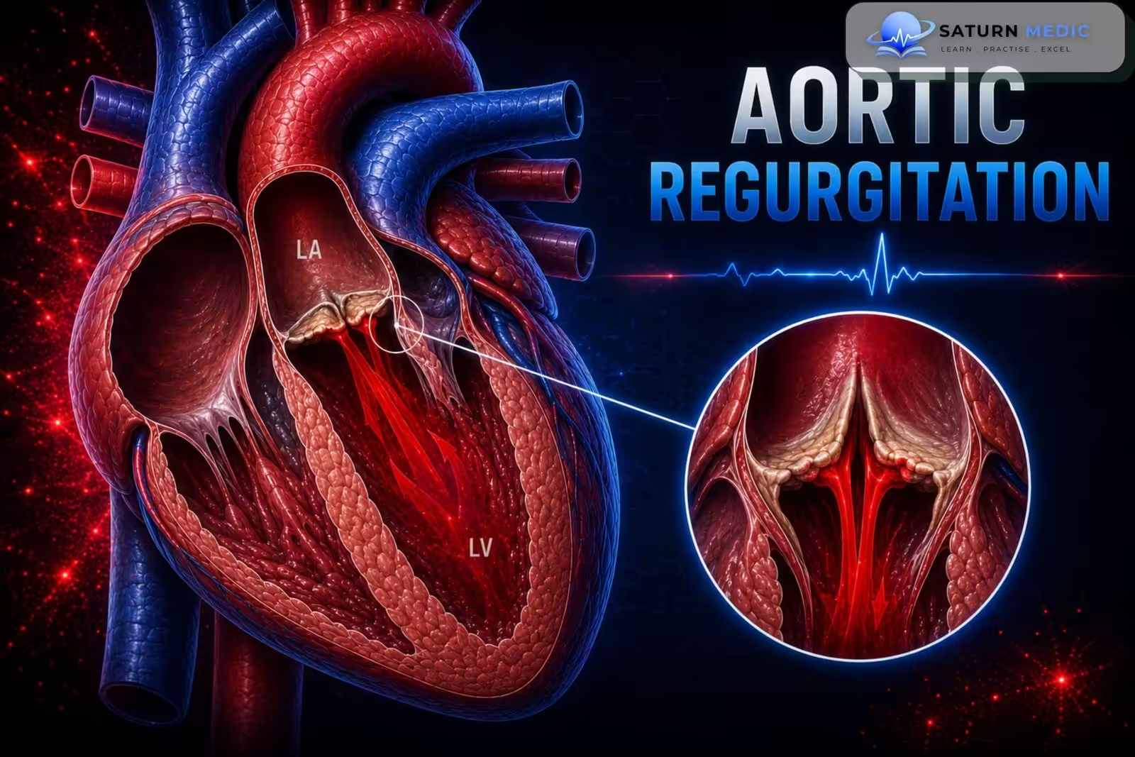
Aortic regurgitation (AR), commonly known as a leaky aortic valve, is a condition in which the aortic valve does not close completely, allowing blood to flow backward from the aorta into the left ventricle during diastole
Etiology & Risk Factors
-Acute Aortic Regurgitation
- Infective endocarditis (most common valvular cause)
- Ascending aortic dissection (most common aortic cause)
- Chest trauma
- Iatrogenic complications (e.g., after TAVR or balloon valvuloplasty)
Chronic Aortic Regurgitation
- Congenital bicuspid aortic valve (most common cause in young adults in high-income countries)
- Calcific aortic valve disease (most common cause in older adults in high-income countries)
- Rheumatic heart disease (most common cause in lower-income countries)
- Aortic root dilation due to:
- Connective tissue disorders (e.g., Marfan syndrome, Ehlers-Danlos syndrome)
- Chronic hypertension
- Aortitis (e.g., tertiary syphilis)
- Thoracic aortic aneurysm
-Risk Factors
- Congenital bicuspid aortic valve
- Calcific aortic valve disease
- Rheumatic heart disease
- Chronic hypertension
- Connective tissue disorders (e.g., Marfan syndrome, Ehlers-Danlos syndrome)
- Thoracic aortic aneurysm
- Infective endocarditis
- Aortic dissection
Pathophysiology
- Aortic valve fails to close completely → blood flows backward from the aorta into the left ventricle (LV) during diastole.
- This causes volume overload of the LV, leading to:
- Increased systolic blood pressure
- Decreased diastolic blood pressure
- Widened pulse pressure (water hammer pulse)
Acute Aortic regurgitation
- The LV cannot dilate quickly enough to accommodate the regurgitant blood.
- LV end-diastolic pressure rises rapidly → blood backs up into the lungs → pulmonary edema and dyspnea.
- Severe cases can cause decreased cardiac output, cardiogenic shock, and myocardial ischemia.
Chronic Aortic regurgitation
- Initially, the LV compensates by increasing stroke volume to maintain cardiac output.
- Over time, persistent volume overload causes LV enlargement (dilation) and eccentric hypertrophy.
- Eventually, LV systolic dysfunction develops, leading to left-sided heart failure.
Flow summary:
Incomplete aortic valve closure → Backflow into LV during diastole → LV volume overload → LV dilation & eccentric hypertrophy → LV dysfunction → Left heart failure (chronic)
Clinical Presentation
-Acute Aortic Regurgitation
Symptoms
- Sudden severe dyspnea
- Pulmonary edema
- Rapid heart failure/cardiac decompensation
- Symptoms of the underlying cause:
- Fever (infective endocarditis)
- Chest pain (aortic dissection)
Auscultation
- Soft S1
- Soft, short early diastolic murmur
-Chronic Aortic Regurgitation
Symptoms
- May be asymptomatic for years
- Palpitations
- Exertional dyspnea
- Angina
- Orthopnea
- Easy fatigability
- Syncope
Signs
- Widened pulse pressure
- Water hammer pulse (bounding pulse)
Auscultation
- High-pitched, blowing, decrescendo early diastolic murmur (best heard along the left sternal border/Erb point in valvular AR)
- S3 heart sound
- Austin Flint murmur (in severe AR)
Key features to remember
- Wide pulse pressure + Water hammer pulse
- High-pitched early diastolic murmur
- Chronic AR can remain asymptomatic for many years before symptoms of left-sided heart failure develop.
History Taking
- “Do you get short of breath, especially when walking or exercising?”
- “Do you have chest pain or discomfort?”
- “Do you notice your heart racing or pounding (palpitations)?”
- “Do you feel unusually tired or fatigued?”
- “Do you feel short of breath when lying flat or wake up at night short of breath?”
- “Have you ever fainted or felt dizzy?”
- “When did your symptoms start? Were they sudden or gradual?”
- “Have you had a recent fever or heart infection?” (suggestive of infective endocarditis)
- “Have you had sudden severe chest pain?” (suggestive of aortic dissection)
- “Have you ever been told you have a heart murmur or valve disease?”
- “Do you have a history of rheumatic fever, high blood pressure, or a connective tissue disorder (e.g., Marfan syndrome)?”
Aortic Stenosis Vs Aortic Regurgitation
Valvular Heart Disease · Aortic Stenosis vs Aortic Regurgitation
| Parameter | Aortic Stenosis | Aortic Regurgitation |
|---|---|---|
| Pathology | Narrowed aortic valve orifice → LV must generate high pressure to eject blood (pressure overload) | Incompetent aortic valve → blood regurgitates back into LV during diastole (volume overload) |
| LV Response | Concentric hypertrophy — wall thickens, cavity size normal or reduced | Eccentric hypertrophy — wall thickens AND cavity dilates (Laplace law) |
| Onset | Usually chronic & insidious (years of compensated disease before symptoms) | Can be chronic (insidious) or acute (endocarditis, aortic dissection — surgical emergency) |
| Classic Triad / Symptoms |
Syncope Angina Dyspnea
SAD triad — appear in this order as severity progresses. Syncope on exertion is a red flag.
|
Exertional dyspnea Palpitations Angina
Long asymptomatic phase. Symptoms indicate LV decompensation. Acute AR: flash pulmonary edema.
|
| Pulse | Pulsus parvus et tardus — small volume, slow-rising, delayed peak | Corrigan's (water-hammer) pulse — large volume, bounding, rapid rise and collapse |
| Blood Pressure | Narrow pulse pressure (<40 mmHg); systolic BP may be low in severe AS | Wide pulse pressure (>60–80 mmHg); high systolic, low diastolic |
| Apex Beat | Sustained (heaving), non-displaced — pressure-loaded LV | Displaced laterally & downward, hyperdynamic — volume-loaded dilated LV |
| Murmur Type | Ejection systolic (crescendo-decrescendo) — heard best at aortic area; radiates to carotids | Early diastolic decrescendo — heard best at left sternal border, leaning forward in expiration |
| Additional Murmurs | S4 (stiff LV); paradoxically split S2 (severe); ejection click (bicuspid valve) | Austin Flint murmur (low-pitched mid-diastolic rumble at apex); S3 (dilated LV) |
| ECG Findings | LVH with strain pattern (ST depression & T-wave inversion in V5–V6, I, aVL); LV strain | LVH (voltage criteria); broad notched P (if LA enlarges); LV volume overload pattern |
| CXR Findings | Normal heart size (concentric LVH); calcified aortic valve; post-stenotic aortic dilatation | Cardiomegaly (LV enlargement); prominent aorta; pulmonary edema if decompensated |
| Echo / Severity | Valve area: mild >1.5 cm², moderate 1.0–1.5 cm², severe <1.0 cm²; mean gradient severe >40 mmHg | Regurgitant fraction: mild <30%, moderate 30–49%, severe ≥50%; LV end-systolic diameter |
| Medical Management | No effective medical therapy to slow progression. Manage comorbidities (HTN, AF). Avoid vasodilators in severe AS. | Vasodilators (nifedipine, ACEi/ARB) to reduce afterload & preload in chronic AR. Bridge to surgery. |
| Surgical Indications | Severe AS + symptoms; severe AS + EF <50%; severe AS undergoing other cardiac surgery | Severe AR + symptoms; severe AR + EF <50%; severe AR + LV ESD >50 mm |
| Intervention Options | SAVR TAVR Balloon valvuloplasty (palliation only) | SAVR Valve repair (if feasible) — TAVR emerging but less established |
| Prognosis | Untreated symptomatic severe AS: 50% mortality at 2–3 yrs. Rapid decline after symptom onset. | Chronic: long compensated phase. Once symptomatic: 10–20% annual mortality without surgery. |
Radiation: Both carotids — key distinguishing feature
Character: Harsh, rasping; peaks in mid-systole; later peak = more severe
Maneuvers: Louder on squatting (increased preload); softer on standing/Valsalva
S2: Soft or absent A2; paradoxical splitting in severe AS
Extras: Ejection click (bicuspid valve); S4 (non-compliant LV)
Position: Patient leaning forward, end-expiration — brings aorta closer to chest wall
Character: Blowing, high-pitched; begins immediately after S2
Maneuvers: Louder on squatting & handgrip (increased afterload)
Austin Flint murmur: Low-pitched mid-diastolic rumble at apex — regurgitant jet impinges on anterior mitral leaflet
Extras: S3 (dilated LV); wide pulse pressure
Investigations
- Transthoracic echocardiography (TTE) – Gold standard for diagnosing Aortic regurgitation, identifying the cause, assessing severity, and evaluating left ventricular function.
- Transesophageal echocardiography (TEE) – Used if TTE is inconclusive, for preoperative planning, or if aortic dissection or infective endocarditis is suspected.
- ECG – May show left ventricular hypertrophy (LVH) in chronic Aortic regurgitation ; also helps assess for arrhythmias or ischemia.
- Chest X-ray – May show left ventricular enlargement, pulmonary edema (acute AR), or a prominent aortic root.
- Laboratory tests
- Blood cultures if infective endocarditis is suspected.
- BNP/NT-proBNP may help assess disease severity.
- CT angiography (CTA) chest – Preferred if aortic dissection is suspected in acute AR.
- Cardiac MRI – Used when echocardiography is inadequate or findings are inconclusive.
- Cardiac catheterization/coronary angiography – Used in selected patients when noninvasive tests are inconclusive or before surgery.
Diagnosis
Aortic Regurgitation · Diagnosis & Severity
| Parameter | Mild | Moderate | Severe |
|---|---|---|---|
| Vena contracta | < 0.3 cm | 0.3–0.6 cm | > 0.6 cm |
| Jet width / LVOT width | < 25% | 25–64% | ≥ 65% |
| Pressure half-time | > 500 ms | 200–500 ms | < 200 ms |
| Regurgitant volume | < 30 mL/beat | 30–59 mL/beat | ≥ 60 mL/beat |
| Regurgitant fraction | < 30% | 30–49% | ≥ 50% |
| EROA | < 0.10 cm² | 0.10–0.29 cm² | ≥ 0.30 cm² |
| Descending aortic flow | No / brief reversal | Intermediate | Holodiastolic reversal |
Management
1. Acute Aortic Regurgitation (Emergency)
- Urgent cardiology and cardiothoracic surgery consultation
- Urgent surgical aortic valve replacement (AVR) is the definitive treatment.
- Stabilize the patient before surgery:
- Treat cardiogenic shock (e.g., with dobutamine or dopamine)
- Reduce afterload for acute heart failure (e.g., nitroprusside)
- Treat the underlying cause (e.g., infective endocarditis or aortic dissection).
- Avoid delaying surgery for medical treatment alone.
- Avoid beta blockers unless the AR is caused by aortic dissection.
- Intra-aortic balloon pump (IABP) is contraindicated because it increases the amount of regurgitation.
2. Chronic Aortic Regurgitation
Asymptomatic patients
- Regular follow-up with serial echocardiography.
- Screen for and treat cardiovascular risk factors.
- Treat hypertension (vasodilators such as ACE inhibitors or ARBs may be preferred).
- Treat heart failure if present.
Symptomatic or severe Aortic regurgitation
- Surgical aortic valve replacement (AVR) is the standard treatment.
3. Indications for Surgery
- Acute severe Aortic regurgitation
- Symptomatic chronic severe Aortic regurgitation
- Asymptomatic severe Aortic regurgitation with:
- LVEF ≤ 55%
- Consider if LV end-systolic diameter (LVESD) > 50 mm
- Patient already undergoing cardiac surgery for another reason
4. Follow-up
- Regular echocardiography to monitor progression:
- Mild AR: every 3–5 years
- Moderate AR: every 1–2 years
- Severe asymptomatic AR: every 6–12 months
Complications
- Left ventricular dilation
- Left ventricular systolic dysfunction
- Left-sided heart failure
- Pulmonary edema (especially in acute Aortic regurgitation)
- Cardiogenic shock (severe acute Aortic regurgitation)
Prognosis
- Asymptomatic patients with normal LV function: Progression to symptoms or LV dysfunction is < 6% per year.
- Asymptomatic patients with reduced ejection fraction: Progression to symptoms is > 25% per year.
- Symptomatic patients: Mortality is > 10% per year and may reach 25% per year if left untreated
Key Points / Clinical Pearls
- Aortic Regurgitation= leaky aortic valve → blood flows back into the LV during diastole.
- Can be acute or chronic.
- Classic murmur: High-pitched, blowing early diastolic decrescendo murmur.
- Classic sign: Wide pulse pressure with a water hammer pulse.
- Gold standard diagnosis: Transthoracic echocardiography (TTE).
- Definitive treatment of Aortic regurgitation : Aortic valve replacement.
- Acute severe Aortic regurgitation is a medical emergency requiring urgent surgery.
- Patibandla S, Heaton J, Azzam JS. National Center for Biotechnology Information (NIH). Aortic Insufficiency, StatPearls.
- Otto CM, Nishimura RA, Bonow RO, et al. 2020 ACC/AHA Guideline for the Management of Patients With Valvular Heart Disease. Circulation. 2021;143:e72-e227. PMID: 33972115.
- Aortic Regurgitation: An Updated Review of Etiologies, Diagnostic Strategies, and Clinical Management. PMC12230845.
- MedlinePlus, National Library of Medicine (NIH). Aortic Regurgitation: Medical Encyclopedia.
- National Center for Biotechnology Information (NIH). Aortic Valve Disease, StatPearls.