

Clinical Subject Page
Cardiac Tamponade & Effusion
Also called
Fluid around the heart
ICD-10
I31.3
Specialty
Cardiology
Onset
Acute or Chronic
Reviewed
June 2026
On This Page
-
OverviewOverview
-
Etiology & Risk FactorsEtiology & Risk Factors
-
PathophysiologyPathophysiology
-
Clinical PresentationClinical Presentation
-
History TakingHistory Taking
-
InvestigationsInvestigations
-
DiagnosisDiagnosis
-
ManagementManagement
-
ComplicationsComplications
-
PrognosisPrognosis
-
Key Points / Clinical PearlsKey Points / Clinical Pearls
Overview
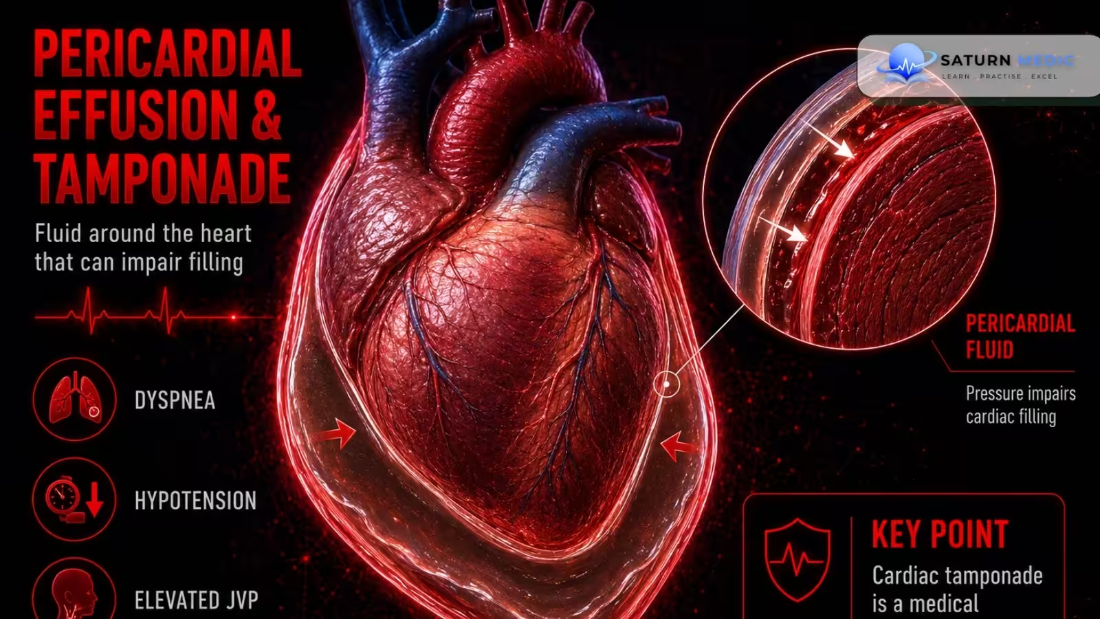
Pericardial effusion is the accumulation of fluid in the pericardial space (between the visceral and parietal pericardium). The fluid may be: Serous (commonly idiopathic) Bloody (hemopericardium) (e.g., after trauma or aortic dissection) The pericardium has limited ability to stretch: Chronic effusion: The pericardium gradually adapts and accommodates more fluid. Acute effusion: Even a small amount of rapidly accumulating fluid can markedly increase intrapericardial pressure.
Etiology & Risk Factors
Hemopericardium (blood in the pericardial space)
- Cardiac wall rupture (e.g., after myocardial infarction)
- Chest trauma
- Aortic dissection
- Cardiac surgery (valve surgery, CABG)
Serous/Serosanguinous effusion
- Idiopathic
- Acute pericarditis (viral most common; also fungal, tuberculous, bacterial)
- Malignancy
- Postpericardiotomy syndrome
- Uremia
- Autoimmune disorders
- Hypothyroidism
- Right heart failure
Pathophysiology
Cardiac Tamponade
Pericardial fluid accumulation leads to:
Pericardial fluid ↑
→ Intrapericardial pressure ↑
→ Compression of the heart (especially the thin-walled right ventricle)
→ Interventricular septum shifts toward the left ventricle
→ Reduced systemic venous return (preload)
→ Reduced ventricular filling during diastole
→ Stroke volume ↓
→ Cardiac output ↓
→ Venous congestion
→ Equalization of end-diastolic pressures in all four cardiac chambers
Clinical Presentation
Pericardial Effusion
Often asymptomatic initially.
Cardiac Tamponade
Typical findings include:
- Hypotension
- Tachycardia
- Jugular venous congestion (raised JVP)
- Pulsus paradoxus
Tamponade is a medical emergency because of reduced cardiac output.
History Taking
Ask about:
- Recent chest trauma
- Recent myocardial infarction
- Cardiac surgery
- Symptoms of acute pericarditis
- History of malignancy
- Kidney disease (uremia)
- Autoimmune disease
- Hypothyroidism
- Symptoms suggesting infection (including tuberculosis)
Investigations
First-line imaging
Echocardiography (TTE) is the most important diagnostic test.
Typical finding:
- Anechoic (fluid-filled) pericardial space
Laboratory tests
- CBC
- CRP
- ESR
- Troponin
- Creatine kinase
- BMP (especially BUN for uremia)
Additional investigations (based on suspected cause)
- Blood cultures
- TB testing (QuantiFERON, HIV test, chest X-ray)
- Renal ultrasound
- Autoimmune tests (ANA, RF, ACPA, ANCA)
- Thyroid function tests
- CT/PET for malignancy
- CT angiography for suspected aortic dissection
- FAST scan or trauma CT in trauma
Pericardial Fluid Analysis
Indicated when the cause is unclear.
Evaluate:
- WBC count
- RBC count
- Gram stain and culture
- Cytology
- Acid-fast bacilli
- Glucose
- Protein
- LDH
Fluid appearance may suggest:
- Transudate: Heart failure, renal failure, hypoalbuminemia
- Exudate: Infection, inflammation, malignancy, autoimmune disease
- Hemorrhagic: Cardiac rupture, aortic dissection, surgery, TB, malignancy
- Purulent: Bacterial infection or tuberculosis
Diagnosis
Diagnostic approach
- Hemodynamically unstable patients: Do not delay treatment for extensive investigations.
- Stable patients: Confirm diagnosis with echocardiography.
- Laboratory studies and pericardial fluid analysis help determine the underlying cause.
In unstable patients or those in cardiac arrest with suspected tamponade, pericardiocentesis should not be delayed for diagnostic confirmation.
Management
Management depends on hemodynamic stability.
Unstable patients (cardiac tamponade)
Urgent drainage of the pericardial fluid by:
- Pericardiocentesis
- Surgical drainage (when indicated)
Stable patients
Treat the underlying cause of the effusion.
Complications
- Cardiac tamponade
- Severe reduction in cardiac output
- Hemodynamic collapse if untreated
Prognosis
Prognosis depends on:
- Hemodynamic stability
- Speed of diagnosis
- Underlying cause
- Prompt drainage when tamponade is present
Rapid recognition and treatment of tamponade are critical because it is life-threatening.
Key Points / Clinical Pearls
- Pericardial effusion = fluid in the pericardial sac.
- Acute effusions are more likely than chronic effusions to cause tamponade because the pericardium cannot rapidly stretch.
- Cardiac tamponade causes:
- Hypotension
- Tachycardia
- Jugular venous congestion
- Pulsus paradoxus
- Echocardiography is the key diagnostic investigation.
- Do not delay pericardiocentesis in unstable patients with suspected tamponade.
- Stable patients should undergo evaluation and treatment of the underlying etiology.
- Willner DA, Goyal A, Grigorova Y, Sharma S, Kiel J. National Center for Biotechnology Information (NIH). Pericardial Effusion, StatPearls.
- National Center for Biotechnology Information (NIH). Cardiac Tamponade, StatPearls.
- National Center for Biotechnology Information (NIH). Pulsus Paradoxus, StatPearls.
- National Center for Biotechnology Information (NIH). Pericardiocentesis, StatPearls.
- Jain CC, Reddy YNV. Pericardial Effusions: Perspective of the Acute Cardiac Care Physician. Eur Heart J Acute Cardiovasc Care. 2023;12:467-474. PMID: 37170756.