

Clinical Subject Page
Supraventricular Tachycardia (SVT)
ICD-10
Specialty
Onset
Reviewed
-
OverviewOverview
-
Etiology & Risk FactorsEtiology & Risk Factors
-
PathophysiologyPathophysiology
-
Clinical PresentationClinical Presentation
-
History TakingHistory Taking
-
Types of SVTTypes of SVT
-
InvestigationsInvestigations
-
DiagnosisDiagnosis
-
ManagementManagement
-
ComplicationsComplications
-
PrognosisPrognosis
-
Key Points / Clinical PearlsKey Points / Clinical Pearls
Overview
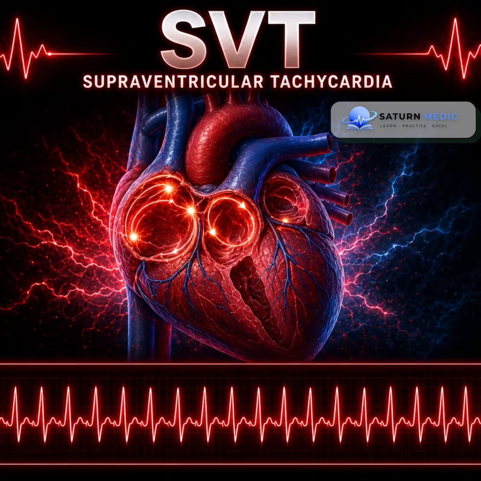
Supraventricular tachycardia (SVT) is a group of rapid heart rhythm disorders that originate above the ventricles, involving the atria or the atrioventricular (AV) node. It is caused by abnormalities in the heart’s electrical conduction or pacemaker activity, most commonly due to reentry circuits.
Etiology & Risk Factors
Supraventicular Tachycardia (SVT) is caused by abnormal electrical impulse formation or conduction above the ventricles. The main mechanisms are:
- Reentry circuits (most common)
- Atrioventricular nodal reentrant tachycardia (AVNRT)
- Atrioventricular reciprocating tachycardia (AVRT)
- Abnormal or ectopic pacemaker activity
- Focal atrial tachycardia (FAT)
- Multifocal atrial tachycardia (MAT)
- Junctional tachycardia
Risk Factors For Supraventricular Tachycardia (SVT)
- Underlying structural heart disease
- Family history of Supraventricular Tachycardia (SVT) or sudden cardiac death
- Use of certain medications (e.g., albuterol, theophylline, methylprednisolone)
- Illicit drug use (e.g., amphetamines, cocaine)
- Hypertension
- Cardiomyopathy
- Myocardial infarction
- Infection
- Alcohol poisoning
- Reentry circuits (most common)
Pathophysiology
- Reentry (most common)
- An electrical impulse repeatedly travels in a continuous loop, causing ongoing rapid stimulation of the heart.
- Occurs in:
- AVNRT: Reentry circuit forms between the fast and slow pathways within the AV node.
- AVRT: Reentry circuit forms between the AV node and an accessory pathway (e.g., Bundle of Kent in WPW syndrome).
- Abnormal automaticity (ectopic pacemaker activity)
- An abnormal focus in the atria or AV junction generates rapid electrical impulses independently of the sinoatrial (SA) node.
- Seen in focal atrial tachycardia and junctional tachycardia.
Summary Flow
Abnormal electrical impulse (reentry or ectopic focus) → Rapid atrial activation → Rapid conduction through the AV node to the ventricles → Supraventricular tachycardia (rapid heart rate) → Reduced ventricular filling time → Symptoms such as palpitations, dizziness, dyspnea, chest pain, or syncope.
- Reentry (most common)
Clinical Presentation
Symptoms of Supraventricular Tachycardia (SVT)
Sudden onset of palpitations (most common)
Fatigue
Chest pain or chest discomfort
Shortness of breath (dyspnea)
Dizziness or lightheadedness (presyncope)
Syncope (more common in older adults)
Diaphoresis (excessive sweating)
Characteristic Features of Supraventricular Tachycardia (SVT)
Symptoms usually begin and end abruptly.
Some patients with AVNRT may experience:
Neck pounding or “shirt flapping”
Urinary urgency or increased urination after an episode
Signs of Unstable Supraventicular Tachycardia (SVT)
Hypotension
Acute pulmonary edema
Severe chest pain
Altered mental status
History Taking
- “Are you feeling short of breath?”
- “Is your breathing worse when lying flat?”
- “Do you have chest pain or pressure?”
- “Have you noticed dizziness or fainting?”
- “Do you feel your heart racing (palpitations)?”
- “Have you had swelling in your legs or abdomen?”
- “Have you had a recent viral illness or pericarditis?”
- “Have you recently had heart surgery or a chest injury?”
- “Do you have kidney disease, cancer, thyroid disease, or an autoimmune condition?”
- “Have you had fever or recent infection?”
- “When did your symptoms start? Did they come on suddenly or gradually?”
Types of Supraventricular Tachycardia (SVT)
Cardiology · Supraventricular Tachycardia (SVT) — Classification & Features
in most SVTs
narrow complex (usual)
~60% of all cases
risk of sudden death in AF
Atypical (fast-slow, slow-slow)
Antidromic (wide QRS) ~5%
Atypical (non-CTI)
Inappropriate sinus tachycardia (IST)
Automatic, triggered, or micro-re-entry
Associated with COPD, hypoxia
Post-cardiac surgery; digitalis toxicity
Technically an SVT but classified separately
EADs — QT prolongation
Typical (slow-fast) ~90%: Antegrade conduction via slow pathway, retrograde via fast pathway. P wave buried in or just after QRS.
Atypical (fast-slow): Antegrade via fast, retrograde via slow — long RP interval; P before next QRS.
QRS Narrow (<120 ms) unless aberrancy
P wave Typical: buried in QRS or pseudo-R’ in V1 / pseudo-S in inf leads — RP < 70 ms
P wave Atypical (fast-slow): long RP > PR
Axis Retrograde P: negative in II, III, aVF
Onset Abrupt; initiated by PAC
Orthodromic AVRT (~95%): Antegrade via AV node (normal, narrow QRS), retrograde via AP. Most common. Normal QRS unless aberrancy.
Antidromic AVRT (~5%): Antegrade via AP (pre-excited, wide QRS), retrograde via AV node. Wide complex — can mimic VT. Seen in WPW.
Delta wave Slurred QRS upstroke — slow initial ventricular activation via AP
QRS Wide (≥120 ms) — fusion of AP + AV node activation
ST/T Discordant (secondary changes)
QRS Narrow (no delta wave — AP used retrogradely)
P wave Retrograde, RP 70–110 ms — longer than AVNRT; P in ST segment
ST Depression common (rate-related)
AV node is a bystander — not part of the circuit. AV block can occur during AT without terminating the tachycardia (key diagnostic feature).
QRS Narrow (unless aberrancy or pre-excitation)
P wave Different morphology from sinus P — shape reveals origin. Long RP > PR (P before QRS or mid-cycle). Isoelectric baseline between P waves
AV block Can occur without terminating AT — pathognomonic of AT
Onset Gradual warm-up (automatic) or abrupt (re-entry)
Atypical flutter: Clockwise (reverse typical), left atrial flutter, or scar-related re-entry (post-ablation, post-surgical). Variable rate and morphology.
Ventricular rate ~150 bpm (2:1 block — most common); 100 (3:1); 75 (4:1)
Flutter waves Sawtooth pattern — negative in II, III, aVF (typical); no isoelectric baseline
V1 Positive flutter waves — upright
QRS Narrow; regular if fixed AV block ratio; irregular if variable
Cause: COPD, hypoxia, electrolyte disturbance, theophylline toxicity, sepsis. Treat the underlying cause. Verapamil or metoprolol may help. Adenosine ineffective (not re-entrant). NOT cardioverted.
P waves ≥3 morphologies, isoelectric baseline
QRS Narrow; variable PR intervals
Key feature: P-wave morphology identical to sinus P wave — only clue is abrupt start/stop and slightly faster rate (100–150 bpm).
Responds to vagal maneuvers and adenosine. Treated with beta-blockers, CCBs, or ablation.
P wave Identical to sinus P — key diagnostic trap
Onset Abrupt (distinguishes from sinus tach)
Causes: Post-cardiac surgery (esp. pediatric), digitalis toxicity, myocarditis, inferior MI, electrolyte disturbance.
AV dissociation may occur. Retrograde P waves (if present) buried in or after QRS. Does NOT respond to adenosine (automatic, not re-entrant). Treat the cause; amiodarone if needed.
P wave Retrograde or AV dissociation
QRS Narrow; gradual onset (warm-up)
| SVT Type | Rate (bpm) | P Wave | RP Interval | QRS | Adenosine | Distinguishing Clue |
|---|---|---|---|---|---|---|
| Typical AVNRT | 150–250 | Buried in QRS; pseudo-R’ V1 / pseudo-S inf | <70 ms (shortest) | Narrow | Terminates | Pseudo-R’ in V1 — absent in sinus rhythm |
| Atypical AVNRT | 150–250 | Before QRS; negative inf leads | Long RP > PR | Narrow | Terminates | Long RP — mimics AT; EPS differentiates |
| Orthodromic AVRT | 150–250 | In ST segment; retrograde, negative inf | 70–110 ms | Narrow | Terminates | Delta wave on sinus ECG (WPW); RP longer than AVNRT |
| Antidromic AVRT | 150–250 | Retrograde after QRS | Variable | Wide (pre-excited) | Terminates (caution) | Fully pre-excited wide QRS; mimics VT |
| Focal AT | 100–250 | Different morphology from sinus; long RP | Long RP > PR | Narrow | AV block only — AT continues | Continues despite AV block; P morphology different from sinus |
| Atrial Flutter | Atrial ~300; Ventricular ~150 | Sawtooth — no isoelectric baseline | N/A | Narrow | Unmasks flutter waves | Ventricular rate 150 = flutter until proven otherwise |
| MAT | >100, irregular | ≥3 morphologies; isoelectric baseline | Variable | Narrow | No effect | Irregular + multiple P morphologies; associated with COPD |
| SANRT | 100–150 | Identical to sinus P | Normal | Narrow | Terminates | Abrupt onset/offset; P wave = sinus P (main trap) |
| Junctional Tachycardia | 70–130 | Retrograde or AV dissociation | Short or AV dissoc. | Narrow | No effect | Post-surgery; warm-up; AV dissociation possible |
Investigations
1. 12-Lead ECG – First-line Investigation
Confirms the diagnosis
Typical findings:
Regular tachycardia
Heart rate >100/min (usually >150/min)
Narrow QRS complex (<120 ms) (may be wide with aberrant conduction)
2. Ambulatory ECG Monitoring
Holter monitor (24–48 hours)
Event recorder
Implantable loop recorder (for infrequent episodes)
3. Echocardiography (TTE)
Evaluates structural heart disease
Assesses cardiac function and heart failure
Initial imaging test of choice
4. Laboratory Tests
CBC (anemia, infection)
BMP (electrolyte abnormalities)
TSH (hyperthyroidism or hypothyroidism)
5. Electrophysiology (EP) Study
Gold standard for identifying the mechanism of Supraventricular Tachycardia (SVT)
Used when the diagnosis is uncertain or before catheter ablation
6. Additional Tests (When Indicated)
Exercise tolerance testing (e.g., suspected WPW syndrome or catecholamine-dependent arrhythmias)
Cardiac stress testing in patients with atherosclerotic cardiovascular disease (ASCVD) risk factors
Diagnosis
Diagnostic Approach
- Confirm the rhythm with a 12-lead ECG in all patients with suspected Supraventricular Tachycardia (SVT).
- Investigate the underlying cause with transthoracic echocardiography (TTE) and laboratory tests.
- If episodes are intermittent, use ambulatory ECG monitoring (Holter monitor or event recorder).
- Consider an electrophysiology (EP) study if the diagnosis is uncertain or catheter ablation is planned.
ECG Findings (Diagnostic)
- Regular tachycardia
- Heart rate >100/min (typically >150/min)
- Narrow QRS complex (<120 ms) (may be wide with aberrant conduction)
Management of Supraventricular Tachycardia (SVT)
1. Initial Management
Assess hemodynamic stability (ABCDE).
Continuous ECG monitoring.
Keep a defibrillator available.
2. Unstable Patient
Immediate synchronized electrical cardioversion.
3. Stable Patient
Step 1: Vagal maneuvers (first-line)
Modified Valsalva maneuver
Carotid sinus massage (only if no carotid bruit)
Diving reflex (selected cases)
Step 2: If Supraventricular Tachycardia (SVT) persists
IV adenosine (first-line if no contraindications)
Step 3: If adenosine is ineffective or contraindicated
Verapamil or diltiazem
OR β-blocker (e.g., metoprolol or esmolol)
Step 4: If Supraventricular Tachycardia (SVT) still persists
Antiarrhythmic drug (e.g., amiodarone or ibutilide)
OR synchronized electrical cardioversion
4. Long-Term Management
Catheter ablation (often curative and first-line for recurrent symptomatic Supraventricular Tachycardia SVT)
Medications if ablation is not suitable:
Beta-blockers
Verapamil or diltiazem
Complications of Supraventricular Tachycardia (SVT)
- Hemodynamic instability (especially if the tachycardia is prolonged or very rapid)
- Syncope
- Heart failure or worsening of existing heart failure
- Tachycardia-induced cardiomyopathy (with persistent or recurrent Supraventricular Tachycardia SVT)
- Sudden cardiac death (rare, mainly in patients with high-risk accessory pathways such as WPW syndrome)
Prognosis of Supraventricular Tachycardia (SVT)
- Most patients have a good prognosis with appropriate treatment.
- Many Supraventricular Tachycardia (SVT) are self-limiting or respond well to vagal maneuvers, medications, or electrical cardioversion.
- Catheter ablation can provide curative treatment for many patients (e.g., AVNRT and AVRT).
- Prognosis depends on the type of SVT, the presence of underlying heart disease, and whether high-risk accessory pathways (e.g., WPW syndrome) are present.
Key Points / Clinical Pearls of Supraventricular Tachycardia (SVT)
- Supraventricular Tachycardia (SVT) is a rapid heart rhythm originating above the ventricles.
- Most common type: AVNRT.
- Diagnosis: 12-lead ECG (Holter monitor if episodes are intermittent).
- First-line treatment in stable patients: Vagal maneuvers → IV adenosine.
- Unstable patients: Immediate synchronized electrical cardioversion.
- Definitive treatment: Catheter ablation for recurrent symptomatic SVT.
- Avoid AV nodal blockers in wide-complex tachycardia or when WPW syndrome is suspected unless the diagnosis is established.
- If the origin of a wide-complex tachycardia is uncertain, treat it as ventricular tachycardia.
- National Center for Biotechnology Information (NIH). Supraventricular Tachycardia, StatPearls.
- Page RL, Joglar JA, Caldwell MA, et al. 2015 ACC/AHA/HRS Guideline for the Management of Adult Patients With Supraventricular Tachycardia. J Am Coll Cardiol. 2016;67:e27-e115. PMID: 26409259.
- Sarkar A, Maria S. National Center for Biotechnology Information (NIH). Electrophysiology Lab Maneuvers to Differentiate Supraventricular Tachycardia, StatPearls.
- MedlinePlus, National Library of Medicine (NIH). Supraventricular Tachycardia: Medical Encyclopedia.
- National Center for Biotechnology Information (NIH). Catheter Ablation, StatPearls.