

Clinical Subject Page
Ventricular Tachycardia (VT)
ICD-10
Specialty
Onset
Reviewed
On This Page
-
OverviewOverview
-
Etiology & Risk FactorsEtiology & Risk Factors
-
PathophysiologyPathophysiology
-
Clinical PresentationClinical Presentation
-
History TakingHistory Taking
-
Types of VTTypes of VT
-
InvestigationsInvestigations
-
DiagnosisDiagnosis
-
ManagementManagement
-
ComplicationsComplications
-
PrognosisPrognosis
-
Key Points / Clinical PearlsKey Points / Clinical Pearls
Overview
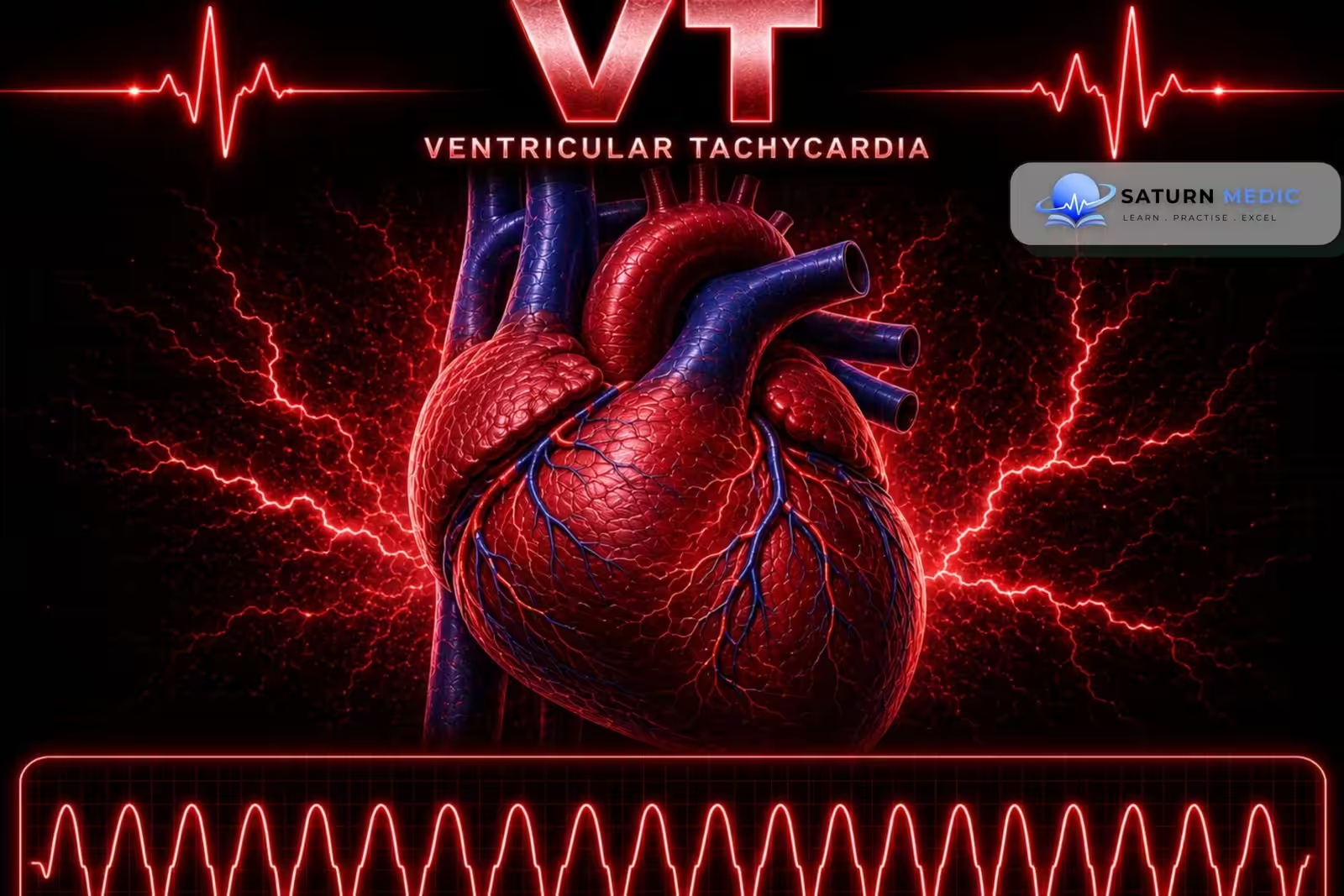
Ventricular tachycardia (VT) is a rapid heart rhythm that originates in the ventricles (the lower chambers of the heart). It is a potentially life-threatening arrhythmia that can reduce the heart’s ability to pump blood effectively and may progress to ventricular fibrillation or sudden cardiac death if not treated promptly.
Etiology & Risk Factors
Etiology
Structural Heart Disease
Previous myocardial infarction (most common cause of sustained monomorphic Ventricular Tachycardia VT )
Ischemic heart disease
Heart failure
Dilated cardiomyopathy
Hypertrophic cardiomyopathy
Arrhythmogenic right ventricular cardiomyopathy (ARVC)
Myocarditis
Cardiac sarcoidosis
Acute Cardiac Causes
Acute myocardial ischemia or infarction
Acute heart failure
Myocarditis
Electrolyte & Metabolic Causes
Hypokalemia
Hypomagnesemia
Severe hypoxemia
Acidosis
Drugs & Toxins
QT-prolonging drugs
Antiarrhythmic drugs
Digoxin toxicity
Stimulants
Inherited Electrical Disorders
Long QT syndrome
Brugada syndrome
Catecholaminergic polymorphic Ventricular Tachycardia VT (CPVT)
Risk Factors For Ventricular Tachycardia (VT)
Previous MI
Reduced left ventricular ejection fraction
Heart failure
Structural heart disease
Previous ventricular arrhythmia
Electrolyte abnormalities
QT prolongation
Family history of sudden cardiac death
Pathophysiology
- Ventricular Tachycardia (VT) results from abnormal myocardial automaticity, electrical conduction, or ventricular repolarization.
- Monomorphic VT
- Caused by:
- Reentry circuit around myocardial scar tissue (most common)
- Increased automaticity (e.g., idiopathic VT)
- Produces wide QRS complexes (≥120 ms).
- Caused by:
- Polymorphic VT
- Caused by abnormal ventricular repolarization, such as:
- Long QT syndrome
- Drug toxicity
- Electrolyte abnormalities
- Caused by abnormal ventricular repolarization, such as:
- Rapid ventricular rhythm causes poor ventricular filling during diastole → decreased cardiac output → hemodynamic compromise.
Simple Flow
Abnormal ventricular electrical activity → Rapid ventricular contractions → Reduced ventricular filling → ↓ Cardiac output → Hypotension, syncope, shock
Clinical Presentation
Common Symptoms
Palpitations
Chest pain or chest pressure
Shortness of breath (dyspnea)
Dizziness
Syncope (fainting)
Signs
Regular tachycardia (HR ≥100/min)
Hypotension
Cardiogenic shock
Loss of consciousness
Cardiac arrest (severe cases)
History Taking
- “Do you feel your heart racing or pounding?”
- “Did the palpitations start suddenly?”
- “Have you had chest pain or chest pressure?”
- “Are you feeling short of breath?”
- “Have you felt dizzy or lightheaded?”
- “Have you fainted or nearly fainted?”
- “Did the symptoms start during exercise or emotional stress?”
- “Do you have a history of a heart attack, heart failure, or cardiomyopathy?”
- “Are you taking any medications that can affect the heart rhythm?”
- “Have you ever been told you have low potassium, low magnesium, or a long QT interval?”
Types of Ventricular Tachycardia (VT)
Ventricular Tachycardia · Types & Classification
Unstable: Synchronised DC cardioversion
Amiodarone IV
Unstable / pulseless: Defibrillation
Correct QT-prolonging drugs & electrolytes
Avoid class Ia/III antiarrhythmics — worsen QT
Catheter ablation if recurrent or ICD shocks
β-blockers for underlying CM
Acquired: remove offending drug; correct electrolytes; pacing if bradycardia-dependent
Investigations
1. 12-Lead ECG – First-line Investigation
Confirms VT
Findings:
Wide QRS complex (>120 ms)
Heart rate ≥100/min (usually 150–200/min)
Regular rhythm (usually)
AV dissociation
Fusion beats or capture beats
2. Laboratory Tests
BMP (electrolytes and renal function)
Serum magnesium
Troponin (if myocardial ischemia is suspected)
BNP/NT-proBNP
Toxicology screen
Drug levels (e.g., digoxin, lithium)
Genetic testing (if inherited arrhythmia is suspected)
3. Imaging
Transthoracic echocardiography (TTE) – first-line imaging
Cardiac MRI (myocardial scar or infiltrative disease)
Cardiac CT (if MRI is contraindicated)
4. Additional Tests
Holter monitor/Event recorder (intermittent VT)
Exercise stress test (exercise-induced VT)
Coronary angiography (suspected myocardial infarction or coronary artery disease)
Electrophysiology (EP) study (to identify the arrhythmia mechanism and before catheter ablation)
Diagnosis
Diagnostic Approach
Obtain a 12-lead ECG in all patients with suspected VT.
If the patient is unstable, presume VT and treat immediately—do not delay treatment for further investigations.
After stabilization, investigate the underlying cause with laboratory tests and cardiac imaging.
Diagnostic ECG Findings
≥3 consecutive wide QRS complexes (>120 ms)
Heart rate ≥100/min (commonly 150–200/min)
Usually regular rhythm
AV dissociation
Fusion beats or capture beats
Management of Ventricular Tachycardia (VT)
1. Initial Management
Assess hemodynamic stability.
Ongoing or sustained Ventricular Tachycardia (VT) :
Stabilize with electrical cardioversion or antiarrhythmic medication.
After stabilization, evaluate and treat the underlying cause.
2. Treat Reversible Causes
Correct electrolyte abnormalities (e.g., potassium and magnesium).
Stop QT-prolonging drugs.
Treat myocardial ischemia if present.
Treat drug toxicity (e.g., digoxin toxicity).
3. Long-Term Management
β-blockers – first-line therapy to reduce the risk of sudden cardiac death.
Amiodarone (often combined with a β-blocker for recurrent VT).
Consider other antiarrhythmic drugs when appropriate.
4. Device Therapy
Implantable cardioverter-defibrillator (AICD/ICD)
Prevents sudden cardiac death.
Used in patients with recurrent VT or those at high risk.
5. Catheter Ablation
Potentially curative treatment.
Indicated for:
Recurrent VT despite optimal therapy.
Intolerance to antiarrhythmic drugs.
Patient preference.
Complications
- Progression to ventricular fibrillation (VF)
- Sudden cardiac death
- Arrhythmia-induced cardiomyopathy
Prognosis
- Generally good with appropriate treatment.
- Catheter ablation has better success and lower recurrence than in atrial fibrillation.
- Many patients respond well to rhythm control, but atrial flutter frequently progresses to atrial fibrillation, so long-term follow-up is important.
Key Points / Clinical Pearls
- Ventricular Tachycardia (VT) is a life-threatening arrhythmia that originates in the ventricles.
- ECG hallmark: Wide QRS complex (>120 ms) with a heart rate ≥100/min.
- Sustained VT is a medical emergency.
- Most common cause: Myocardial infarction (MI) and structural heart disease.
- Symptoms: Palpitations, chest pain, dyspnea, dizziness, and syncope.
- Diagnosis: 12-lead ECG.
- Unstable Ventricular Tachycardia (VT): Immediate synchronized cardioversion (or defibrillation if pulseless).
- Treat reversible causes: Correct electrolyte abnormalities, stop QT-prolonging drugs, and treat myocardial ischemia.
- Long-term treatment: β-blockers, ICD (AICD), and catheter ablation when indicated.
- Major complications: Ventricular fibrillation and sudden cardiac death.
- Lovik K, Ahmed I. National Center for Biotechnology Information (NIH). Ventricular Tachycardia, StatPearls.
- National Center for Biotechnology Information (NIH). Pulseless Ventricular Tachycardia, StatPearls.
- Al-Khatib SM, Stevenson WG, Ackerman MJ, et al. 2017 AHA/ACC/HRS Guideline for Management of Patients With Ventricular Arrhythmias and the Prevention of Sudden Cardiac Death. J Am Coll Cardiol. 2018;72:e91-e220. PMID: 29097296.
- Napolitano C, Mazzanti A, Bloise R, Priori SG. National Center for Biotechnology Information (NIH). Catecholaminergic Polymorphic Ventricular Tachycardia, GeneReviews.
- MedlinePlus, National Library of Medicine (NIH). Ventricular Tachycardia: Medical Encyclopedia.