

Clinical Subject Page
Aortic Dissection
ICD-10
Specialty
Onset
Reviewed
-
OverviewOverview
-
Etiology & Risk FactorsEtiology & Risk Factors
-
PathophysiologyPathophysiology
-
Clinical PresentationClinical Presentation
-
History TakingHistory Taking
-
Act ImmediatelyAct Immediately
-
InvestigationsInvestigations
-
DiagnosisDiagnosis
-
ManagementManagement
-
ComplicationsComplications
-
PrognosisPrognosis
-
Key Points / Clinical PearlsKey Points / Clinical Pearls
Overview
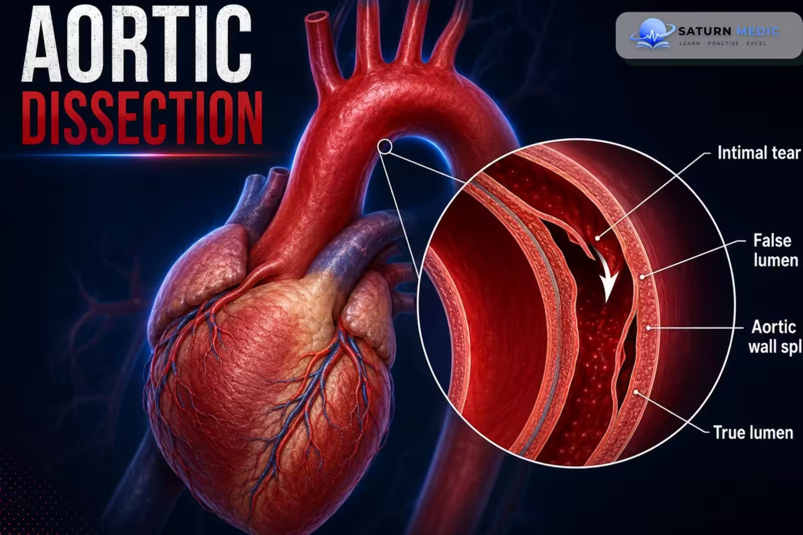
Aortic dissection is a life-threatening condition in which an intimal tear allows blood to split the layers of the aortic wall, creating a false lumen that can impair organ perfusion or rupture.
Etiology & Risk Factors
Etiology
-
Chronic hypertension (most common)
-
Connective tissue disorders (Marfan, Ehlers-Danlos)
-
Bicuspid aortic valve
-
Aortic aneurysm
-
Atherosclerosis
-
Iatrogenic injury (catheterization, surgery)
-
Blunt chest trauma
-
Pregnancy (rare)
Risk Factors
-
-
-
Male sex
-
Age >60 years
-
Long-standing hypertension
-
Smoking
-
Family history of aortic disease
-
Cocaine or stimulant use
-
Known thoracic aortic aneurysm
-
-
Pathophysiology
-
-
-
-
An aortic dissection occurs when a tear develops in the aortic intima, allowing blood to enter the wall of the aorta and separate its layers.
Mechanism
- A transverse tear in the intima (entry tear) occurs.
- Blood enters the media of the aortic wall.
- Blood dissects between the intima and media, creating a false lumen alongside the normal true lumen.
- The hematoma spreads (propagates) along the length of the aorta.
Consequences
- Rising pressure in the aortic wall may cause aortic rupture.
- The false lumen can compress or occlude branch arteries, causing ischemia of organs supplied by:
- Coronary arteries → myocardial ischemia/infarction
- Carotid arteries → stroke
- Renal arteries → acute kidney injury
- Mesenteric arteries → bowel ischemia
- Lower limb arteries → limb ischemia
- Sometimes a second intimal tear (re-entry tear) forms, allowing blood to flow back into the true lumen.
Simple Flow
Intimal tear → Blood enters the media → False lumen forms → Dissection spreads → Compression of branch vessels + risk of aortic rupture → Organ ischemia and life-threatening hemorrhage
-
-
-
Clinical Presentation
Symptoms
-
Sudden severe tearing chest pain
-
Back or interscapular pain
-
Abdominal pain
-
Syncope
-
Dyspnea
-
Neurological deficits
Signs
-
-
Blood pressure difference between arms
-
Pulse deficit
-
New early diastolic murmur (aortic regurgitation)
-
Hypotension or shock
-
Focal neurological deficits
-
History Taking
Key Questions
-
Sudden onset of pain?
-
Chest, back, or abdominal pain?
-
Pain migration?
-
Syncope?
-
Weakness or stroke symptoms?
-
History of hypertension?
-
Connective tissue disorder?
-
Cocaine use?
-
Previous aneurysm or aortic surgery?
Red Flags
-
-
Sudden tearing pain
-
Hypotension
-
Neurological deficit
-
Pulse asymmetry
-
Signs of cardiac tamponade
-
Act Immediately
Aortic Dissection · Immediate Management
| Drug | Dose / Target | Role / Notes |
|---|---|---|
| Esmolol | 500 mcg/kg load, then 50–200 mcg/kg/min infusion | Ultra-short-acting cardioselective beta-blocker; first-line for rapid, titratable HR control. |
| Labetalol | 20 mg IV bolus, then infusion 1–2 mg/min | Combined alpha/beta blockade — controls both HR and BP; useful alternative/adjunct to esmolol. |
| Sodium Nitroprusside | 0.3–0.5 mcg/kg/min, titrate to SBP 100–120 | Potent arterial/venous vasodilator. Only after adequate beta-blockade to avoid reflex tachycardia. |
| Nicardipine | 5 mg/hr, titrate up to 15 mg/hr | Alternative vasodilator to nitroprusside; avoids cyanide toxicity risk, easier to titrate. |
| IV Opioid Analgesia | Morphine/Fentanyl titrated | Pain itself raises catecholamines/HR/BP — adequate analgesia supports haemodynamic control. |
Investigations
- Laboratory
CBC
Renal function
Troponin
D-dimer (may support exclusion in low-risk patients)
ECG
Often normal or nonspecific
May show ischemic changes - Imaging
CT angiography (first-line in stable patients)
Transesophageal echocardiography (unstable patients)
MRI angiography (stable patients) - Chest X-ray
Widened mediastinum
Pleural effusion
Diagnosis
- Clinical suspicion
- CT angiography (preferred)
- TEE or MRI when appropriate
- Classification using Stanford (A/B) or DeBakey systems
Management
Initial Medical Therapy
-
ICU admission
-
IV β-blocker (Esmolol or Labetalol)
-
Target HR <60 bpm
-
IV vasodilator (Nicardipine/Nitroprusside) after β-blockade
-
Adequate pain control (IV opioids)
Definitive Treatment
-
-
-
-
Stanford Type A: Emergency surgical repair
-
Stanford Type B: Medical management unless complications develop; TEVAR for complicated cases
-
-
-
Complications
- Aortic rupture
- Cardiac tamponade
- Acute aortic regurgitation
- Myocardial infarction
- Stroke
- Acute kidney injury
- Mesenteric ischemia
- Limb ischemia
- Death
Prognosis
- Untreated Stanford type A dissection has a very high mortality.
- Early diagnosis and prompt treatment significantly improve survival.
- Long-term follow-up with strict blood pressure control is essential.
Key Points / Clinical Pearls
- Hypertension is the most common risk factor For Aortic Dissection.
- CT angiography is the diagnostic test of choice in stable patients.
- Lower heart rate before lowering blood pressure.
- Stanford Type A = Surgery; Type B = Usually medical management.
- Sudden tearing chest or back pain is the classic presentation.
- Levy D, Sharma S, Farci F, Le JK. National Center for Biotechnology Information (NIH). Aortic Dissection, StatPearls.
- Isselbacher EM, Preventza O, Hamilton Black J 3rd, et al. 2022 ACC/AHA Guideline for the Diagnosis and Management of Aortic Disease. Circulation. 2022;146:e334-e482. PMID: 36334952.
- Vignaraja V, Sharma S, Dindyal S. National Center for Biotechnology Information (NIH). Acute Aortic Syndrome, StatPearls.
- Hussain K, Huerter M. National Center for Biotechnology Information (NIH). ACC/AHA Guidelines for Aortic Disease, StatPearls.
- MedlinePlus, National Library of Medicine (NIH). Aortic Dissection: Medical Encyclopedia.