

Clinical Subject Page
Deep Vein Thrombosis (DVT)
ICD-10
Specialty
Onset
Reviewed
-
OverviewOverview
-
Etiology & Risk FactorsEtiology & Risk Factors
-
PathophysiologyPathophysiology
-
Clinical PresentationClinical Presentation
-
History TakingHistory Taking
-
Physical ExaminationPhysical Examination
-
InvestigationsInvestigations
-
Diagnosis (Well's Score)Diagnosis (Well's Score)
-
ManagementManagement
-
ComplicationsComplications
-
PrognosisPrognosis
-
Key Points / Clinical PearlsKey Points / Clinical Pearls
Overview
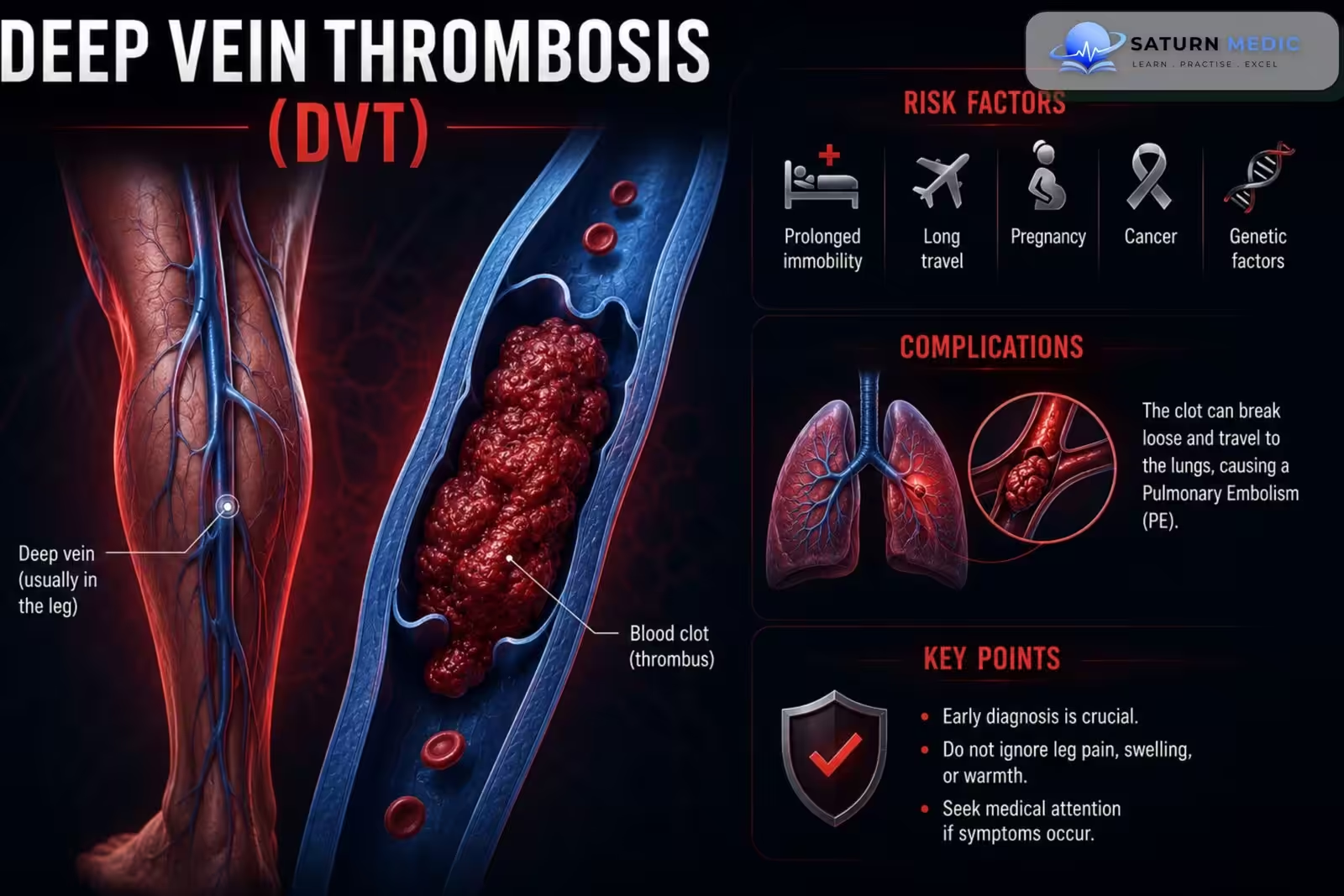
Deep vein thrombosis (DVT) is the formation of a thrombus within the deep venous system, most commonly in the lower extremities, which may obstruct venous blood flow and lead to pulmonary embolism (PE).
Etiology & Risk Factors
Etiology for Deep Vein Thrombosis (DVT)
Venous stasis
Endothelial injury
Hypercoagulable states (Virchow’s triad)
Recent surgery
Trauma
Malignancy
Pregnancy/postpartum
Estrogen therapy (OCP/HRT)
Inherited thrombophilia
Risk Factors for Deep Vein Thrombosis (DVT)
Previous Deep Vein Thrombosis (DVT)/PE
Prolonged immobilization
Hospitalization
Active cancer
Obesity
Older age
Smoking
Heart failure
Stroke with paralysis
Long-distance travel
Pathophysiology
-Deep Vein Thrombosis (DVT) develops due to Virchow’s triad, which includes three major factors that promote thrombus formation.
Virchow’s Triad
- Hypercoagulability
- Increased tendency for blood to clot
- Examples: thrombophilia, pregnancy, oral contraceptive use
- Endothelial Damage
- Injury to the vein wall exposes subendothelial collagen
- Activates the coagulation cascade and platelet adhesion
- Causes include trauma and inflammation
- Venous Stasis
- Slow or stagnant venous blood flow
- Causes include:
- Prolonged immobilization
- Hospitalization/bed rest
- Long flights
- Varicose veins
- External compression of veins
Simple Flow
Virchow’s triad (Hypercoagulability + Endothelial injury + Venous stasis) → Thrombus forms in deep vein → Venous obstruction → Impaired venous return → Leg swelling, pain, warmth → Risk of pulmonary embolism if clot embolizes
Clinical Presentation
Symptoms
Unilateral leg swelling
Calf pain or tenderness
Leg heaviness
Warmth
Redness
Dilated superficial veins
Signs
Unilateral pitting edema
Calf tenderness
Increased calf circumference
Erythema
Warm affected limb
History Taking
Key Questions
When did the leg swelling begin?
Is the swelling unilateral?
Recent surgery or hospitalization?
Prolonged immobility or long travel?
Previous DVT or PE?
Active cancer?
Pregnancy or postpartum?
Use of oral contraceptives or hormone therapy?
Chest pain or shortness of breath?
Red Flags for Deep Vein Thrombosis (DVT)
Sudden dyspnea
Chest pain
Hemoptysis
Syncope
Massive leg swelling (phlegmasia)
Physical Examination
Inspection
Unilateral leg swelling (edema)
Redness (erythema)
Dilated superficial veins (may be present)
Palpation
Warm, tender calf or thigh
Calf tenderness along the course of the deep veins
Increased calf circumference compared with the opposite leg
Vascular Examination
Compare both legs for:
Swelling
Temperature
Tenderness
Circumference
Severe Deep Vein Thrombosis (DVT) (Emergency)
Marked swelling
Cold, cyanotic, pulseless limb suggests phlegmasia cerulea dolens, which requires emergency treatment.
Investigations
Laboratory
D-dimer
CBC
Coagulation profile
Renal function
Thrombophilia testing (selected patients)
Imaging
Compression duplex ultrasonography (first-line test)
CT or MR venography (selected cases)
Additional Tests
Wells score for Deep Vein Thrombosis (DVT) probability
CT pulmonary angiography if PE is suspected
Diagnosis (Well's Score)
Wells Score · Deep Vein Thrombosis (DVT) Probability
| Clinical Criterion | Points |
|---|---|
| Active cancer (treatment ongoing, within 6 months, or palliative) | +1 |
| Paralysis, paresis, or recent plaster immobilisation of the lower extremity | +1 |
| Recently bedridden ≥3 days, or major surgery within 12 weeks requiring general/regional anaesthesia | +1 |
| Localised tenderness along the deep venous system | +1 |
| Entire leg swollen | +1 |
| Calf swelling ≥3 cm larger than asymptomatic leg (measured 10 cm below tibial tuberosity) | +1 |
| Pitting oedema confined to the symptomatic leg | +1 |
| Collateral superficial veins (non-varicose) | +1 |
| Previously documented DVT | +1 |
| Alternative diagnosis at least as likely as DVT (e.g. cellulitis, Baker's cyst, muscle strain) | −2 |
| Total Score | Sum points |
Management
General Measures
Early ambulation
Leg elevation
Graduated compression stockings (selected patients)
Anticoagulation
Direct oral anticoagulants (Apixaban, Rivaroxaban)
Low-molecular-weight heparin (LMWH)
Unfractionated heparin
Warfarin (selected patients)
Advanced Therapy
Catheter-directed thrombolysis (selected extensive DVT)
Mechanical thrombectomy
Inferior vena cava (IVC) filter (if anticoagulation is contraindicated)
Complications
-
- Pulmonary embolism
- Post-thrombotic syndrome
- Recurrent Deep Vein Thrombosis (DVT)
- Chronic venous insufficiency
- Phlegmasia cerulea dolens
Prognosis
- Excellent with prompt anticoagulation.
- Untreated Deep Vein Thrombosis (DVT) significantly increases the risk of pulmonary embolism.
- Some patients develop chronic post-thrombotic syndrome despite treatment.
Key Points / Clinical Pearls
- Virchow’s triad is the foundation of Deep Vein Thrombosis (DVT) pathophysiology.
- Compression duplex ultrasound is the first-line diagnostic test.
- Assess clinical probability using the Wells score.
- DOACs are first-line therapy for most patients.
- Always evaluate for pulmonary embolism in patients with DVT who develop chest pain or dyspnea.
- Early anticoagulation reduces recurrence and mortality.
- National Center for Biotechnology Information (NIH). Deep Venous Thrombosis, StatPearls.
- Kearon C, Akl EA, Ornelas J, et al. Antithrombotic Therapy for VTE Disease: CHEST Guideline and Expert Panel Report. Chest. 2016;149:315-352. PMID: 26867832.
- Wells PS, Anderson DR, Rodger M, et al. Excluding Pulmonary Embolism at the Bedside Without Diagnostic Imaging: Management of Patients With Suspected Pulmonary Embolism Presenting to the Emergency Department by Using a Simple Clinical Model and D-Dimer. Ann Intern Med. 2001;135:98-107. PMID: 11453709.
- MedlinePlus, National Library of Medicine (NIH). Deep Vein Thrombosis: Health Topic.
- Konstantinides SV, Meyer G, Becattini C, et al. 2019 ESC Guidelines for the Diagnosis and Management of Acute Pulmonary Embolism Developed in Collaboration With the European Respiratory Society (ERS). Eur Respir J. 2019;54:1901647. PMID: 31473594.