

Clinical Subject Page
Peripheral Artery Disease (PAD)
ICD-10
Specialty
Onset
Reviewed
-
OverviewOverview
-
Etiology & Risk FactorsEtiology & Risk Factors
-
PathophysiologyPathophysiology
-
Clinical PresentationClinical Presentation
-
History TakingHistory Taking
-
ClassificationClassification
-
Investigations ApproachInvestigations Approach
-
DiagnosisDiagnosis
-
ManagementManagement
-
ComplicationsComplications
-
PrognosisPrognosis
-
Key Points / Clinical PearlsKey Points / Clinical Pearls
Overview
Peripheral artery disease (PAD) is a chronic atherosclerotic disease causing narrowing or occlusion of peripheral arteries, most commonly in the lower extremities, resulting in reduced blood flow and limb ischemia.
Etiology & Risk Factors
Etiology of Peripheral Artery Disease (PAD)
Atherosclerosis (most common)
Diabetes mellitus
Chronic kidney disease
Vasculitis
Thromboembolism
Radiation-induced vascular disease
Buerger disease (thromboangiitis obliterans)
Risk Factors for Peripheral Artery Disease (PAD)
Smoking (strongest modifiable risk factor)
Diabetes mellitus
Hypertension
Hyperlipidemia
Older age
Family history
Chronic kidney disease
Obesity
Pathophysiology
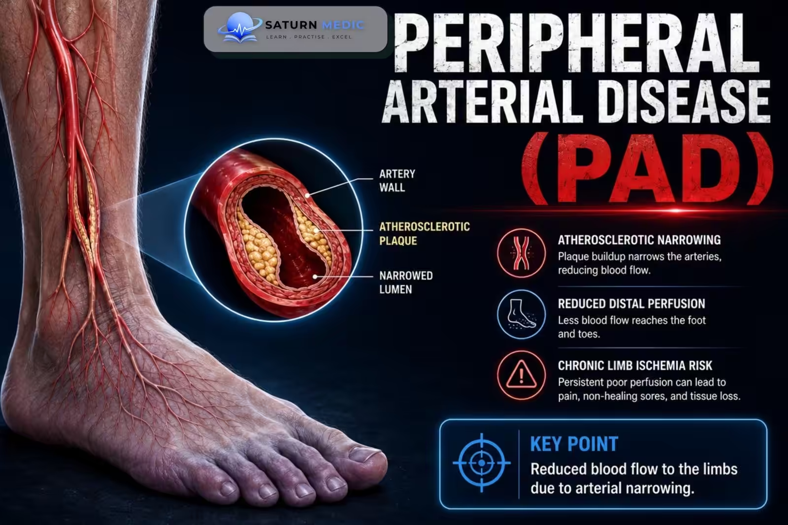
Peripheral artery disease (PAD) is caused by progressive atherosclerosis of the peripheral arteries, leading to reduced blood flow and oxygen delivery to the limbs, especially during exercise.
Mechanism
Atherosclerotic risk factors:
- Smoking
- Diabetes
- Hypertension
- Hyperlipidemia
This causes:
- Endothelial injury
- Atherosclerotic plaque formation
- Progressive arterial narrowing (stenosis)
As the disease progresses:
- Reduced arterial blood flow
- Inadequate oxygen delivery during exercise
- Limb ischemia
- Tissue ischemia at rest in advanced disease
- Non-healing ulcers and gangrene
Simple Flow
Risk factors → Endothelial injury → Atherosclerotic plaque formation → Progressive arterial narrowing → ↓ Limb blood flow → Ischemia → Intermittent claudication → Critical limb ischemia (advanced disease)
Clinical Presentation
Symptoms of Peripheral Artery Disease (PAD)
Intermittent claudication
Leg fatigue during walking
Rest pain (advanced disease)
Non-healing foot ulcers
Cold feet
Numbness
Signs of Peripheral Artery Disease (PAD)
Diminished or absent peripheral pulses
Cool extremity
Pallor on elevation
Dependent rubor
Delayed capillary refill
Hair loss
Shiny skin
Ischemic ulcers
History Taking
Key Questions For Peripheral Artery Disease (PAD)
Leg pain while walking?
How far can you walk before pain starts?
Does pain improve with rest?
Rest pain?
Non-healing wounds?
Smoking history?
Diabetes?
Previous cardiovascular disease?
History of stroke or MI?
Red Flags
Rest pain
Tissue loss
Gangrene
Sudden limb pain
Acute limb ischemia
Classification
Peripheral Arterial Disease (PAD) · Classification
| Rutherford Grade | Category | Clinical Description | Equivalent Fontaine |
|---|---|---|---|
| 0 | 0 | Asymptomatic — no haemodynamically significant disease | Stage I |
| I | 1 | Mild claudication | Stage IIa |
| I | 2 | Moderate claudication | Stage IIa/IIb |
| I | 3 | Severe claudication | Stage IIb |
| II | 4 | Ischaemic rest pain | Stage III |
| III | 5 | Minor tissue loss — non-healing ulcer, focal gangrene with diffuse pedal ischaemia | Stage IV |
| III | 6 | Major tissue loss — extending above transmetatarsal level, functional foot no longer salvageable | Stage IV |
Investigations Approach
Peripheral Arterial Disease · Investigations Approach
| Investigation | Role |
|---|---|
| Ankle-Brachial Index | First-line, bedside, diagnostic and severity marker for chronic PAD; not used in acute limb ischaemia workup |
| Duplex Ultrasound | Non-invasive first-line imaging; localises stenosis/occlusion, assesses flow velocity and vessel patency |
| CT Angiography | Detailed anatomical mapping for revascularisation planning; investigation of choice in acute limb ischaemia |
| MR Angiography | Alternative to CT if contrast/radiation contraindicated; avoid gadolinium in severe renal impairment |
| Digital Subtraction Angiography | Gold standard but invasive; usually reserved for concurrent endovascular intervention |
| Handheld Doppler | Rapid bedside confirmation of arterial signal presence/absence in acute or chronic settings |
Diagnosis
-
-
-
Diagnosis of Peripheral Artery Disease (PAD) is based on:
- Clinical symptoms
- ABI ≤0.90
- Duplex ultrasound
- CT/MR angiography when revascularization is planned
-
-
Management
Lifestyle Modification
Smoking cessation
Supervised exercise therapy
Weight control
Foot care
Medications
Antiplatelet therapy (Aspirin or Clopidogrel)
High-intensity statin
ACE inhibitor/ARB (if indicated)
Cilostazol (for claudication, if no heart failure)
Revascularization
Endovascular angioplasty ± stenting
Surgical bypass
Amputation (non-salvageable limb)
Complications
- Critical limb ischemia
- Non-healing ulcers
- Gangrene
- Limb amputation
- Acute limb ischemia
- Myocardial infarction
- Stroke
Prognosis
- Stable with risk-factor modification and medical therapy.
- Patients with Peripheral Artery Disease (PAD) have a significantly increased risk of cardiovascular events.
- Critical limb ischemia carries a high risk of amputation and mortality.
Key Points / Clinical Pearls
- Smoking is the strongest modifiable risk factor for Peripheral Artery Disease (PAD).
- Atherosclerosis is the most common cause of PAD.
- ABI is the first-line diagnostic test (ABI ≤0.90 confirms PAD).
- Intermittent claudication is the classic presentation.
- All patients require aggressive cardiovascular risk reduction.
- Critical limb ischemia is a vascular emergency requiring urgent revascularization.
- Zemaitis MR, Boll JM, Kato M, Golla MSG. National Center for Biotechnology Information (NIH). Peripheral Arterial Disease, StatPearls.
- Gornik HL, Aronow HD, Goodney PP, et al. 2024 ACC/AHA/AACVPR/APMA/ABC/SCAI/SVM/SVN/SVS/SIR/VESS Guideline for the Management of Lower Extremity Peripheral Artery Disease. Circulation. 2024;149:e1313-e1410. PMID: 38743805.
- MedlinePlus, National Library of Medicine (NIH). Peripheral Arterial Disease: Health Topic.
- National Center for Biotechnology Information (NIH). Ankle-Brachial Index, StatPearls.
- National Center for Biotechnology Information (NIH). Chronic Limb-Threatening Ischemia, StatPearls.