

Clinical Subject Page
Atrial Septal Defect (ASD)
ICD-10
Specialty
Onset
Reviewed
-
OverviewOverview
-
Etiology & Risk FactorsEtiology & Risk Factors
-
PathophysiologyPathophysiology
-
Clinical PresentationClinical Presentation
-
History TakingHistory Taking
-
InvestigationsInvestigations
-
DiagnosisDiagnosis
-
ManagementManagement
-
ComplicationsComplications
-
PrognosisPrognosis
-
Key Points / Clinical PearlsKey Points / Clinical Pearls
Overview
Atrial Septal Defect (ASD) is A defect in the atrial septum caused by impaired growth or excessive resorption of the atrial septum
Etiology & Risk Factors
- – Down syndrome
– Fetal alcohol syndrome
– Intrauterine infections (e.g., TORCH)
– Holt-Oram syndrome (hand-heart syndrome)
– Autosomal dominant
– Characterized by:
– Atrial septal defect
– First-degree heart block
– Upper limb abnormalities (e.g., absent radial bones)
Pathophysiology
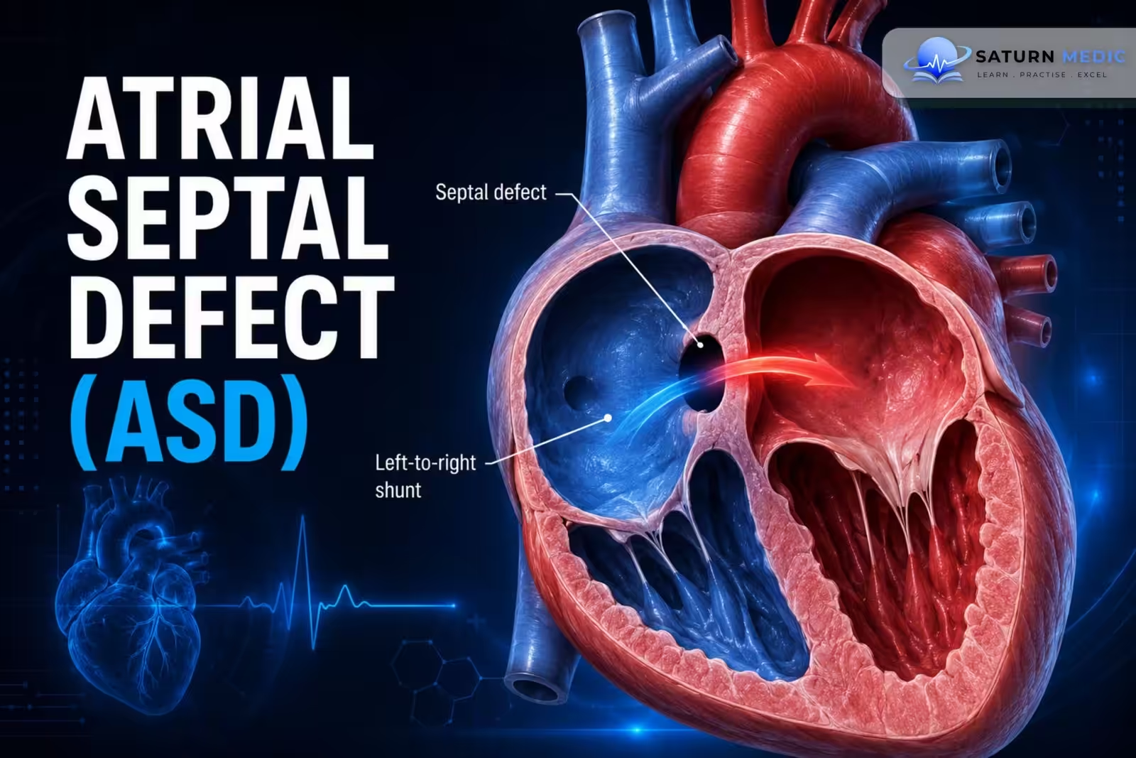
- Impaired growth or excessive resorption of the atrial septum during fetal development creates a defect in the atrial septum.
- This allows oxygenated blood to flow from the left atrium (LA) to the right atrium (RA) because left atrial pressure is higher than right atrial pressure.
- The left-to-right shunt increases:
- Right atrial blood volume
- Right ventricular blood volume
- Pulmonary blood flow
- Small Atrial Septal Defect (ASD) usually cause a low-pressure, low-volume shunt, so patients are often asymptomatic.
- Large Atrial Septal Defect (ASD) can lead to:
- Right heart volume overload
- Pulmonary hypertension
- Supraventricular arrhythmias
- Eisenmenger syndrome (late reversal to right-to-left shunt)
Simple Flow
Defect in atrial septum → Left-to-right shunt (LA → RA) → ↑ Blood flow to RA, RV & lungs → Right heart volume overload → Pulmonary hypertension → Arrhythmias ± Eisenmenger syndrome (late)
Clinical Presentation
- Small Atrial Septal Defect (ASD) :
– Usually asymptomatic
- Medium to Large Atrial Septal Defect (ASD)
- Symptoms often develop in adulthood (around 30–40 years):
- Exertional dyspnea
- Fatigue
- Recurrent respiratory infections
- Palpitations (supraventricular arrhythmias)
- Syncope
- Heart failure symptoms (e.g., peripheral edema)
- Stroke or TIA due to paradoxical embolism
-Auscultation
Mid-systolic ejection murmur at the 2nd left intercostal space
– Fixed, widely split second heart sound (S2)
– Soft mid-diastolic murmur at the lower left sternal border
History Taking
Evaluate for:
– Exertional dyspnea
– Fatigue
– Palpitations
– Recurrent respiratory infections
– Syncope
– Heart failure symptoms
– Previous stroke or TIA (suggesting paradoxical embolism)
Investigations
Echocardiography (Confirmatory Test)
– Transthoracic echocardiography (TTE) with Doppler
– Confirms interatrial communication
– Best seen in apical four-chamber and subcostal views
– Agitated saline study if findings are equivocal
– TEE if TTE is limited or for intraoperative guidanceECG
May show:
– Right ventricular hypertrophy
– Right axis deviation
– P pulmonale
– PR prolongation
– Complete or incomplete right bundle branch block
– Atrial fibrillation/flutter (common in adults)Chest X-ray
– Enlarged right atrium
– Enlarged right ventricle
– Enlarged pulmonary artery
– Increased pulmonary vascular markings
Pulse Oximetry
– At rest and during exercise
– Helps assess magnitude and direction of shunt
Diagnosis
Diagnosis of Atrial Septal Defect (ASD) is confirmed by:
– Transthoracic echocardiography with Doppler (first-line confirmatory investigation)
Supportive findings:
– ECG abnormalities
– Chest X-ray changes
– Cardiac MRI/CT
– Cardiac catheterization when detailed hemodynamics are required
Management
General Principles
– Managed by a congenital cardiologist
– Treat associated conditions:
– Atrial fibrillation
– Tachyarrhythmias
– Pulmonary hypertension
– Small/asymptomatic ASD:
– Clinical follow-up
– Serial echocardiography
– Larger or symptomatic Atrial Septal Defect (ASD) :
– Surgical closure
Indications for Closure
– Large left-to-right shunt (Qp:Qs ≥1.5:1 without pulmonary hypertension)
– Right atrial or ventricular hypertrophy
– Heart failure symptoms
– Previous paradoxical embolism
– Platypnea-orthodeoxia syndrome
Contraindications
– Severe pulmonary hypertension
– Eisenmenger syndrome (right-to-left shunt)
Closure Methods
– Transcatheter closure
– Patch repair
– Open surgical repair- Surgical closure
Indications for Closure
– Large left-to-right shunt (Qp:Qs ≥1.5:1 without pulmonary hypertension)
– Right atrial or ventricular hypertrophy
– Heart failure symptoms
– Previous paradoxical embolism
– Platypnea-orthodeoxia syndrome
Contraindications
– Severe pulmonary hypertension
– Eisenmenger syndrome (right-to-left shunt)
Closure Methods
– Transcatheter closure
– Patch repair
– Open surgical repair
Complications
- Paradoxical embolism leading to ischemic stroke
- Heart failure
Prognosis
- – Up to 40% of ASDs close spontaneously by 5 years of age.
– Larger symptomatic defects generally require closure.
– Surgical repair is contraindicated in patients with Eisenmenger syndrome or right-to-left shunting.
Key Points / Clinical Pearls
- – VSD is the most common congenital heart defect.
– Most defects are membranous.
– Small VSDs produce louder murmurs than large VSDs.
– Classic murmur: harsh holosystolic murmur at the left lower sternal border.
– Confirmatory test: transthoracic echocardiography with Doppler.
– Many small VSDs close spontaneously.
– Close significant defects before irreversible pulmonary hypertension develops.
– Do not close VSDs in patients with Eisenmenger syndrome.
- Menillo AM, Alahmadi MH, Pearson-Shaver AL. National Center for Biotechnology Information (NIH). Atrial Septal Defect, StatPearls.
- 2025 ACC/AHA/HRS/ISACHD/SCAI Guideline for the Management of Adults With Congenital Heart Disease. Circulation. 2025. doi: 10.1161/CIR.0000000000001402.
- Oster M, Bhatt AB, Zaragoza-Macias E, et al. Interventional Therapy Versus Medical Therapy for Secundum Atrial Septal Defect: A Systematic Review. J Am Coll Cardiol. 2019;73:1579-1595. PMID: 30121241.
- MedlinePlus, National Library of Medicine (NIH). Atrial Septal Defect: Medical Encyclopedia.
- Basit H, Wallen TJ, Sergent BN. National Center for Biotechnology Information (NIH). Eisenmenger Syndrome, StatPearls.