

Clinical Subject Page
Varicose Veins
ICD-10
Q25.0
Specialty
Cardiology
Onset
Chronic
Reviewed
July 2026
On This Page
-
OverviewOverview
-
Etiology & Risk FactorsEtiology & Risk Factors
-
PathophysiologyPathophysiology
-
Clinical PresentationClinical Presentation
-
History TakingHistory Taking
-
Physical ExaminationPhysical Examination
-
InvestigationsInvestigations
-
DiagnosisDiagnosis
-
ManagementManagement
-
ComplicationsComplications
-
PrognosisPrognosis
-
Key Points / Clinical PearlsKey Points / Clinical Pearls
Overview
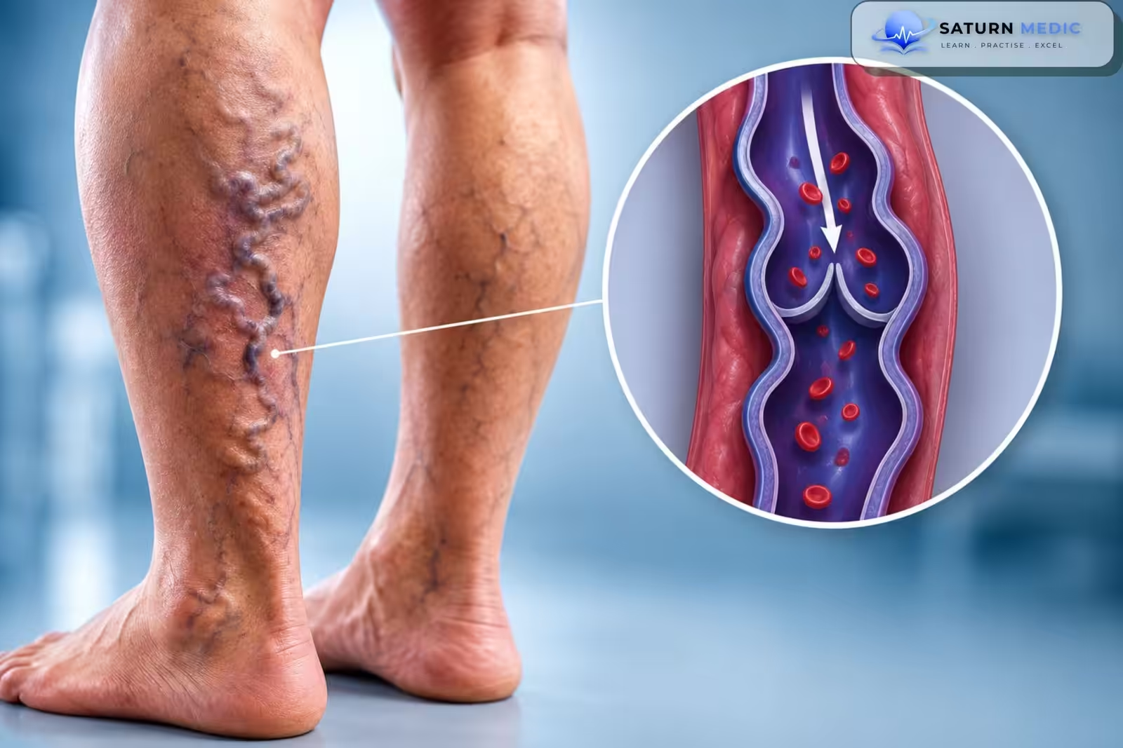
Varicose veins are dilated, tortuous superficial veins caused by venous valve incompetence, leading to venous reflux and chronic venous hypertension, most commonly affecting the lower limbs.
Teaching point
Varicose veins are the most common manifestation of chronic venous disease and result from venous valve failure causing reflux and venous hypertension.
Etiology & Risk Factors
Etiology
- Primary venous valve incompetence (most common)
- Chronic venous insufficiency
- Previous deep vein thrombosis (post-thrombotic syndrome)
- Congenital venous valve abnormalities
- Venous obstruction
Risk Factors
- Increasing age
- Female sex
- Family history
- Pregnancy
- Obesity
- Prolonged standing
- Sedentary lifestyle
- Previous DVT
Pathophysiology
- – During fetal development:
– Anterior and superior deviation of the infundibular septum causes: - Malaligned VSD
- Overriding aorta
- Right ventricular outflow tract obstruction
- Blood flow depends on the severity of RVOTO:
- Large VSD causes equal right and left ventricular pressures.
- Mild RVOTO → predominantly left-to-right shunt → minimal cyanosis.
- Severe RVOTO → right-to-left shunt → deoxygenated blood enters systemic circulation.
- Persistent right-to-left shunting eventually results in right ventricular hypertrophy
Clinical Presentation
Symptoms
Visible enlarged veins
Leg heaviness
Aching pain
Leg fatigue
Swelling
Itching
Night cramps
Signs
Dilated tortuous superficial veins
Ankle edema
Hyperpigmentation
Lipodermatosclerosis
Venous eczema
Venous ulcers (advanced disease)
Important Note
Symptoms typically worsen after prolonged standing and improve with leg elevation or walking.
History Taking
Key Questions
- When did the veins first appear?
- Leg pain or heaviness?
- Symptoms worse after standing?
- Swelling?
- Skin discoloration?
- Previous DVT?
- Pregnancy history?
- Family history?
- Occupation requiring prolonged standing?
Red Flags
- Sudden unilateral swelling
- Severe pain
- Bleeding from varicose veins
- Non-healing ulcer
- Suspected DVT
Physical Examination
Inspection
- Dilated, tortuous superficial veins (>3 mm in diameter)
- Leg swelling (edema), usually starting at the ankle
- Telangiectasias (spider veins)
- Yellow-brown or red-brown skin pigmentation around the medial ankle (hemosiderin deposition)
- Stasis dermatitis (scaly, itchy rash)
- Lipodermatosclerosis (hard, indurated skin of the lower leg)
- Venous ulcers (typically around the medial malleolus in advanced disease)
Palpation
- Assess for:
- Tenderness
- Warmth
- Extent of edema
- Induration from lipodermatosclerosis
Vascular Examination
- Check pedal pulses to exclude coexisting peripheral arterial disease.
- Compare both legs for swelling and skin changes.
Bedside Clinical Tests
- Trendelenburg test: Assesses superficial and perforating vein valve competence.
- Perthes test: Assesses deep venous patency (deep vein function).
Note: These bedside tests may help identify venous insufficiency, but venous duplex ultrasound is required for diagnostic confirmation.
Investigations
Laboratory
- Usually not required
- D-dimer (if DVT suspected)
Imaging
- Duplex venous ultrasound (gold standard)
- Venous reflux
- Valve incompetence
- Excludes DVT
Additional Tests
- CEAP classification
- Venous Clinical Severity Score (VCSS)
Diagnosis
Diagnosis is based on:
- Clinical examination
- Duplex venous ultrasound confirming venous reflux and valve incompetence
- CEAP classification to determine disease severity
Management
Conservative Management
- Compression stockings
- Leg elevation
- Regular exercise
- Weight reduction
- Avoid prolonged standing
Procedures
- Endovenous thermal ablation (laser or radiofrequency)
- Ultrasound-guided foam sclerotherapy
- Ambulatory phlebectomy
- Surgical vein stripping (selected patients)
Complications
- Chronic venous insufficiency
- Venous ulcers
- Superficial thrombophlebitis
- Bleeding
- Skin pigmentation
- Lipodermatosclerosis
- Recurrent varicosities
Prognosis
- Generally excellent with appropriate treatment.
- Symptoms improve significantly after endovenous therapy.
- Recurrence may occur, especially if risk factors persist.
- Untreated disease may progress to chronic venous insufficiency and venous ulceration.
Key Points / Clinical Pearls
- Venous valve incompetence is the primary cause of varicose veins.
- Duplex venous ultrasound is the diagnostic test of choice.
- Compression therapy is the first-line conservative treatment.
- Endovenous thermal ablation is the preferred minimally invasive intervention.
- Symptoms worsen with prolonged standing and improve with leg elevation.
- Venous ulcers are the most significant long-term complication.
- National Center for Biotechnology Information (NIH). Varicose Veins, StatPearls.
- National Center for Biotechnology Information (NIH). Venous Insufficiency, StatPearls.
- National Institute for Health and Care Excellence (NICE). Varicose Veins in the Legs: Guideline Summary.
- MedlinePlus, National Library of Medicine (NIH). Varicose Veins: Health Topic.
- Singh A, Zahra F. National Center for Biotechnology Information (NIH). Chronic Venous Insufficiency, StatPearls.