

Clinical Subject Page
Cardiac Arrest
Also called
ICD-10
Specialty
Onset
Reviewed
-
OverviewOverview
-
Etiology & Risk FactorsEtiology & Risk Factors
-
PathophysiologyPathophysiology
-
Clinical PresentationClinical Presentation
-
Recognition of Cardiac Arrest (Initial Assessment)Recognition of Cardiac Arrest (Initial Assessment)
-
InvestigationsInvestigations
-
DiagnosisDiagnosis
-
ManagementManagement
-
ComplicationsComplications
-
PrognosisPrognosis
-
Key Points / Clinical PearlsKey Points / Clinical Pearls
Overview
Cardiac arrest is the sudden cessation of cardiac function and blood circulation, resulting in loss of consciousness, apnea (or abnormal gasping), and absence of a pulse. It is a medical emergency requiring immediate CPR and, if indicated, defibrillation to improve survival.
Etiology & Risk Factors
Cardiac Causes
Coronary artery disease (most common in adults)
Acute myocardial infarction
Malignant arrhythmias (VF, pulseless VT)
Cardiomyopathy
Severe heart failure
Noncardiac Causes (Reversible Causes)
5 Hs
Hypovolemia
Hypoxia
Hydrogen ion (acidosis)
Hyperkalemia/Hypokalemia
Hypothermia
5 Ts
Tension pneumothorax
Cardiac tamponade
Toxins
Thrombosis (pulmonary embolism)
Thrombosis (coronary)
Other causes include:
Hypoglycemia
Hypocalcemia
Hypomagnesemia
Anaphylaxis
Severe asthma
Pathophysiology
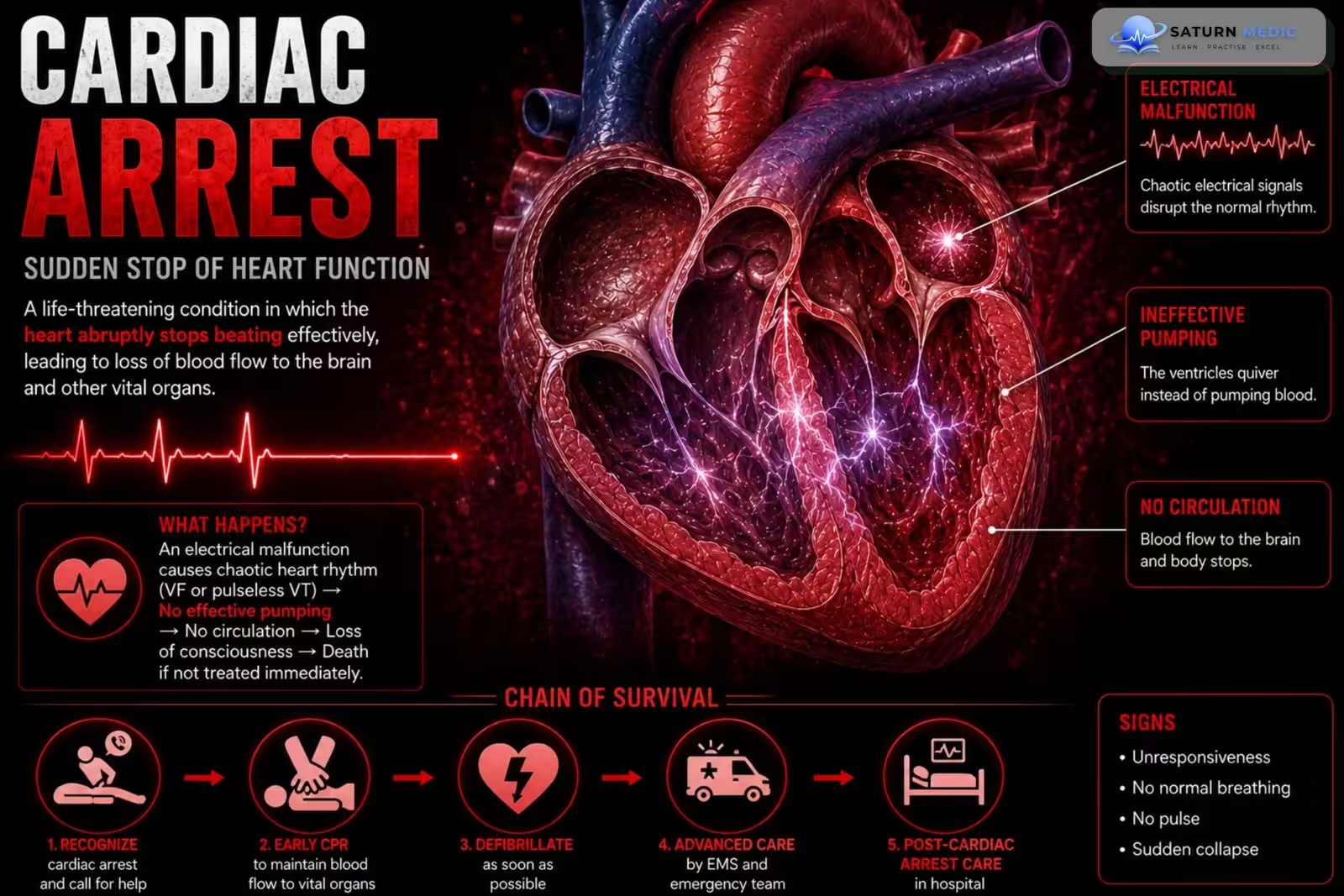
Cardiac arrest occurs when the heart suddenly stops pumping blood effectively, causing cessation of systemic circulation.
Mechanism
- Loss of effective cardiac mechanical activity
- No cardiac output
- No blood flow to vital organs
- Rapid cerebral hypoxia and ischemia
- Irreversible brain injury begins within minutes if circulation is not restored
Simple Flow
Underlying cardiac/noncardiac cause → Heart stops pumping effectively → No cardiac output → No oxygen delivery to organs → Brain ischemia → Organ failure → Death (without prompt CPR/defibrillation)
Clinical Presentation
Symptoms/Signs
- Sudden collapse
- Unresponsiveness
- No normal breathing (or gasping)
- No palpable pulse
Recognition of Cardiac Arrest (Initial Assessment)
Step 1: Ensure Scene Safety
Make sure the environment is safe for you and the patient.
Step 2: Check Responsiveness
Tap the patient’s shoulders.
Shout: “Are you okay?”
If there is no response, suspect cardiac arrest.
Step 3: Call for Help
Activate the emergency response system.
Ask someone to bring an AED/defibrillator immediately.
Step 4: Check Breathing
Look for normal breathing.
Agonal gasps are NOT normal breathing and should be treated as cardiac arrest.
Step 5: Check Pulse (Healthcare Providers)
Check a central pulse (carotid or femoral).
Spend no more than 10 seconds checking.
If no pulse or unsure → start CPR immediately.
Step 6: Start High-Quality CPR
Begin chest compressions immediately.
Attach an AED/defibrillator as soon as it is available.
Signs Suggesting Cardiac Arrest
Sudden collapse
Unresponsiveness
No normal breathing or only gasping
No palpable central pulse
Pale or cyanotic skin
Dilated pupils (late sign)
Investigations
Laboratory
CBC
Electrolytes
Renal & liver function
Troponin
BNP/NT-proBNP
Arterial blood gas
Serum lactate
Coagulation profile
ECG
Acute MI changes
Arrhythmias
Imaging
Echocardiography (first-line)
Chest X-ray
Coronary angiography (if ACS suspected)
Hemodynamic Monitoring
Arterial line
Central venous access
Pulmonary artery catheter (selected patients)
Diagnosis
Diagnosis is based on:
- Clinical evidence of shock
- SBP <90 mmHg (or need for vasopressors)
- Signs of end-organ hypoperfusion
- Elevated lactate
- Echocardiographic evidence of severe cardiac dysfunction
- Identification of the underlying cardiac cause
Management
1. Basic Life Support (BLS)
Recognize cardiac arrest and activate the emergency response system.
Start high-quality CPR immediately.
Apply an AED (Automated External Defibrillator) as soon as available.
Defibrillate immediately if a shockable rhythm is detected.
2. Advanced Cardiac Life Support (ACLS)
Step 1
Continue high-quality CPR.
Attach a cardiac monitor/defibrillator.
Obtain IV/IO access.
Step 2: Identify the Rhythm
A. Shockable Rhythms
Ventricular fibrillation (VF)
Pulseless ventricular tachycardia (pVT)
Management
Immediate defibrillation
Resume CPR for 2 minutes
Epinephrine 1 mg IV/IO after the second unsuccessful shock, then every 3–5 minutes
Amiodarone 300 mg IV/IO (or lidocaine) after the third unsuccessful shock
B. Nonshockable Rhythms
Pulseless electrical activity (PEA)
Asystole
Management
Do NOT defibrillate
Continue CPR
Epinephrine 1 mg IV/IO immediately, then every 3–5 minutes
Search for and treat reversible causes (5 Hs & 5 Ts)
3. Airway Management
Minimize interruptions to CPR.
Consider advanced airway (endotracheal tube or supraglottic airway) if needed.
Confirm placement with continuous waveform capnography.
Once an advanced airway is in place:
Continue continuous chest compressions.
Give 1 breath every 6 seconds.
4. Post-Resuscitation Care (After ROSC)
Secure the airway and optimize oxygenation/ventilation.
Maintain hemodynamic stability.
Consider PCI if a cardiac cause (e.g., STEMI) is suspected.
Temperature control.
Treat seizures if present.
Monitor for organ dysfunction and neurological recovery.
Complications
- Anoxic-ischemic encephalopathy (brain injury) (most important complication)
- Neurological dysfunction
- Ischemic cardiomyopathy
- Acute kidney injury (AKI)
- Shock liver
- Multiorgan dysfunction syndrome (MODS)
- Seizures
- Coma, persistent vegetative state, or brain death (in severe cases)
Prognosis
- Prognosis depends mainly on:
- Time to CPR
- Time to defibrillation (for shockable rhythms)
- Duration of anoxia
- Presence of return of spontaneous circulation (ROSC)
- Early CPR and early defibrillation significantly improve survival and neurological outcomes.
- Anoxia lasting >3–5 minutes is commonly associated with permanent brain injury.
- Patients who achieve ROSC require comprehensive post-resuscitation care to optimize recovery.
Key Points / Clinical Pearls
- Cardiac Arrest is the sudden cessation of effective cardiac mechanical activity, causing loss of consciousness and absence of normal breathing.
- In an adult who is unresponsive and not breathing normally or only gasping, assume cardiac arrest.
- A healthcare professional should check for a pulse for no more than 10 seconds; if no definite pulse is felt, start CPR.
- Activate the emergency response system and obtain an AED/defibrillator immediately.
- Start high-quality CPR immediately.
- Adult CPR uses 30 chest compressions : 2 breaths.
- Compression rate: 100–120/minute.
- Compression depth in adults: 5–6 cm.
- Allow complete chest recoil and minimize interruptions.
- Use an AED/defibrillator as soon as available.
- Shockable rhythms: ventricular fibrillation (VF) and pulseless ventricular tachycardia (pVT).
- Non-shockable rhythms: pulseless electrical activity (PEA) and asystole.
- If the rhythm is shockable → 1 shock → immediately resume CPR for 2 minutes
- National Center for Biotechnology Information (NIH). Cardiopulmonary Arrest in Adults, StatPearls.
- Panchal AR, Bartos JA, Cabañas JG, et al. Part 3: Adult Basic and Advanced Life Support: 2020 American Heart Association Guidelines for CPR and Emergency Cardiovascular Care. Circulation. 2020;142:S366-S468. doi: 10.1161/CIR.0000000000000916.
- National Center for Biotechnology Information (NIH). Advanced Cardiac Life Support (ACLS), StatPearls.
- MedlinePlus, National Library of Medicine (NIH). Sudden Cardiac Death: Medical Encyclopedia.
- National Center for Biotechnology Information (NIH). Pulseless Electrical Activity, StatPearls.