

Clinical Subject Page
Mallory-Weiss Syndrome
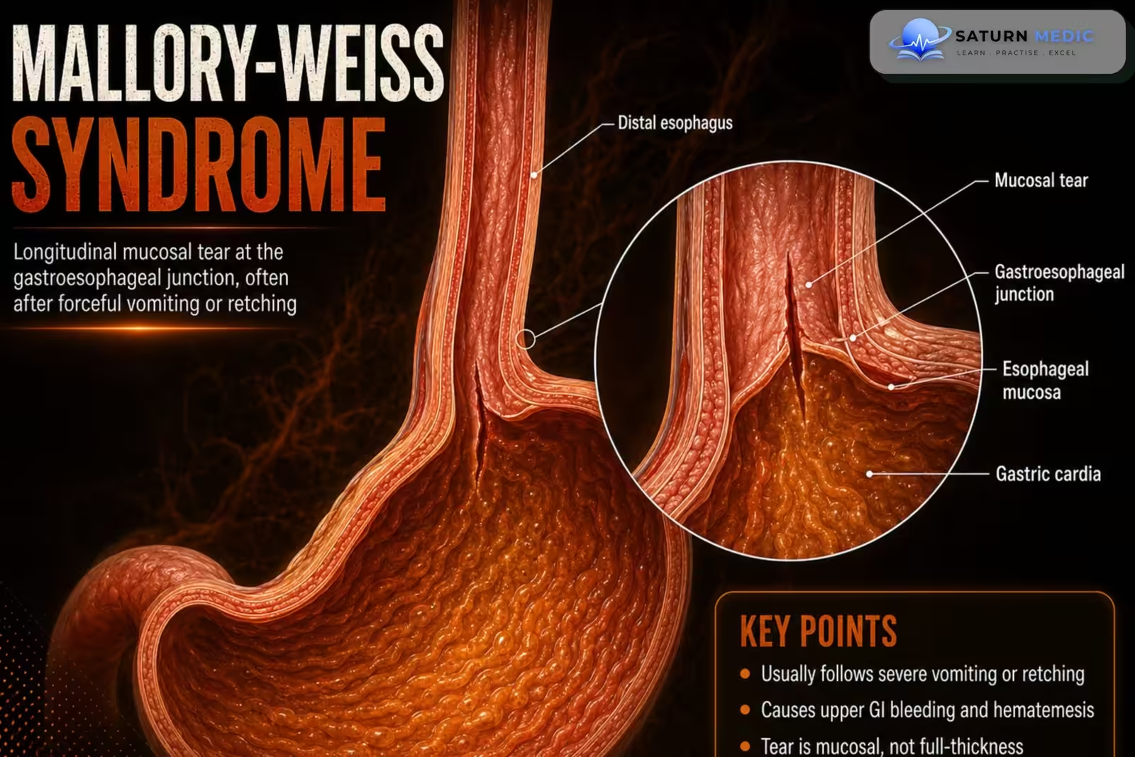
Mallory-Weiss syndrome is an acute upper gastrointestinal (GI) bleeding disorder caused by
mucosal lacerations at the gastroesophageal junction. The tears may also extend above and/or
below the gastroesophageal junction.
Also called
ICD-10
Specialty
Onset
Reviewed
-
OverviewOverview
-
Etiology & Risk FactorsEtiology & Risk Factors
-
PathophysiologyPathophysiology
-
Clinical PresentationClinical Presentation
-
History TakingHistory Taking
-
Physical ExaminationPhysical Examination
-
InvestigationsInvestigations
-
DiagnosisDiagnosis
-
ManagementManagement
-
ComplicationsComplications
-
PrognosisPrognosis
-
Key Points / Clinical PearlsKey Points / Clinical Pearls
Overview
Mallory-Weiss tears most commonly occur after forceful vomiting, especially in patients with gastric
mucosal injury related to heavy alcohol use. Patients usually present with hematemesis following
episodes of retching or vomiting. Most cases resolve with supportive treatment, while persistent
bleeding may require endoscopic hemostasis.
Etiology & Risk Factors
Common Causes
Forceful or repeated vomiting (most common)
Severe retching
Excessive alcohol intake
Persistent coughing
Recurrent hiccups
Seizures
Heavy lifting or straining
Blunt abdominal trauma (rare)
Upper gastrointestinal endoscopy (rare, iatrogenic)
Risk Factors
Heavy alcohol use
Recurrent vomiting (e.g., gastroenteritis)
Eating disorders (e.g., bulimia nervosa)
Pregnancy with hyperemesis gravidarum
Hiatal hernia
Chronic cough
Older age
Conditions causing repeated retching or increased intra-abdominal pressure
Pathophysiology
1. Forceful vomiting causes a sudden increase in pressure across the gastroesophageal junction.
2. Longitudinal mucosal lacerations develop at the gastroesophageal junction.
3. The tears bleed, resulting in upper GI hemorrhage.
4. Most bleeding stops spontaneously, but severe hemorrhage can occur.
Clinical Presentation
–Typical features include:
• Hematemesis
• Epigastric pain
• History of forceful vomiting or retching
–Severe cases:
• Hemodynamic instability due to significant blood loss
History Taking
-
Ask about:
• Recent forceful vomiting or retching
• Hematemesis
• Epigastric pain
• Amount of blood vomited
• Alcohol use
• Previous episodes of GI bleeding
• Symptoms of dizziness or syncope suggesting significant blood loss
Physical Examination
Assess for:
• Signs of upper GI bleeding
• Hemodynamic instability
• Tachycardia
• Hypotension
• Evidence of volume depletion
Investigations
Initial Tests
Complete blood count (CBC)
Blood type and crossmatch (if significant bleeding)
Coagulation profile (PT/INR, aPTT)
Renal function tests and electrolytes
Liver function tests (especially in patients with alcohol-related liver disease)
Diagnostic Investigation
Upper Gastrointestinal Endoscopy (EGD) – Gold Standard
Gold standard for diagnosis
Identifies the mucosal tear at the gastroesophageal junction
Determines whether bleeding is active
Allows immediate endoscopic treatment if needed
Additional Investigations
CT angiography (rarely, if endoscopy is nondiagnostic and bleeding persists)
Catheter angiography for persistent or recurrent bleeding when endoscopic therapy fails
Diagnosis
-
Diagnosis is usually based on:
• Typical history (forceful vomiting followed by hematemesis)
• Confirmation with EGD
Related Topics
Management
1. Initial Stabilization
Assess airway, breathing, and circulation (ABC)
Establish IV access
Fluid resuscitation if needed
Blood transfusion for significant blood loss or hemodynamic instability
2. Medical Therapy
Proton pump inhibitors (PPIs)
Antiemetics to control nausea and prevent further vomiting
Correct any coagulopathy if present
3. Endoscopic Therapy
Indicated for active or ongoing bleeding.
Options include:
Endoscopic hemoclip placement
Endoscopic band ligation
Injection therapy (e.g., epinephrine)
Thermal coagulation
4. Angiographic or Surgical Management
Reserved for patients with:
Persistent bleeding despite endoscopic therapy
Recurrent hemorrhage
Hemodynamic instability
Options:
Angiographic embolization
Surgical repair (rare)
5. Follow-Up
Treat the underlying cause of vomiting or retching
Avoid excessive alcohol consumption
Avoid NSAIDs when possible
Monitor for recurrent bleeding
Complications
• Acute upper GI bleeding
• Hemodynamic instability
• Significant blood loss requiring transfusion
Prognosis
- Most patients recover with supportive care. Bleeding often stops spontaneously, and further
treatment is frequently unnecessary in patients without active bleeding. - Severe hemorrhage
generally responds to endoscopic therapy.
Key Points / Clinical Pearls
• Mallory-Weiss Syndrome is a mucosal laceration at the gastroesophageal junction.
• The classic cause is forceful vomiting or retching.
• Heavy alcohol use is a common associated risk factor.
• Hematemesis and epigastric pain are the typical presenting features.
• EGD is the diagnostic test of choice and can provide therapeutic hemostasis.
• Initial management focuses on hemodynamic stabilization.
• Antiemetics and acid suppression are recommended.
• Angiography is considered if endoscopic treatment fails.
• Surgery is rarely necessary
- Kassama Z, Goosenberg E. National Center for Biotechnology Information (NIH). Mallory-Weiss Syndrome, StatPearls.
- Yin A, Li Y, Jiang Y, Liu J, Luo H. Mallory-Weiss Syndrome: Clinical and Endoscopic Characteristics. Eur J Intern Med. 2012;23:e92-e96. PMID: 22726377.
- MedlinePlus, National Library of Medicine (NIH). Mallory-Weiss Tear: Medical Encyclopedia.
- National Center for Biotechnology Information (NIH). Upper Gastrointestinal Bleeding, StatPearls.
- Kim JW, Kim HS, Byun JW, et al. Predictive Factors of Recurrent Bleeding in Mallory-Weiss Syndrome. Korean J Gastroenterol. PMC5793389.