

Clinical Subject Page
Zenker Diverticulum
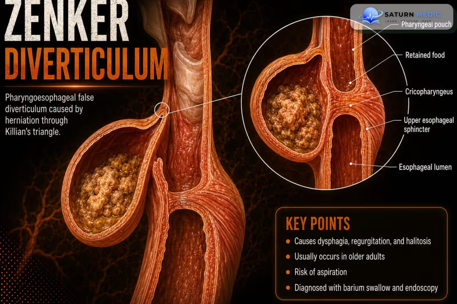
Zenker diverticulum is the most common upper esophageal (pharyngoesophageal) diverticulum. It
is a false (pulsion) diverticulum that arises through the Killian triangle, a weak area in the posterior
wall of the hypopharynx.
Also called
ICD-10
Specialty
Onset
Reviewed
-
OverviewOverview
-
Etiology & Risk FactorsEtiology & Risk Factors
-
PathophysiologyPathophysiology
-
Clinical PresentationClinical Presentation
-
History TakingHistory Taking
-
Physical ExaminationPhysical Examination
-
InvestigationsInvestigations
-
DiagnosisDiagnosis
-
ManagementManagement
-
ComplicationsComplications
-
PrognosisPrognosis
-
Key Points / Clinical PearlsKey Points / Clinical Pearls
Overview
Zenker diverticulum results from increased intraluminal pressure causing the mucosa and
submucosa to herniate through the Killian triangle. It typically presents with regurgitation and
halitosis, and is diagnosed with a modified barium swallow. Symptomatic patients are usually
treated surgically or endoscopically
Etiology & Risk Factors
Common Causes
Cricopharyngeal muscle dysfunction (most common)
Impaired relaxation of the upper esophageal sphincter
Increased hypopharyngeal pressure during swallowing
Age-related degeneration of pharyngeal muscles
Esophageal motility disorders (less common)
Risk Factors
Age > 60 years
Male sex
Cricopharyngeal dysfunction
Esophageal motility disorders
Chronic swallowing dysfunction (dysphagia)
Connective tissue degeneration with aging
Pathophysiology
1. Increased intraluminal pressure develops in the hypopharynx.
2. The mucosa and submucosa herniate through the Killian triangle.
3. A false (pulsion) diverticulum forms.
4. Food collects within the diverticulum, leading to characteristic symptoms.
Clinical Presentation
–Typical symptoms include:
• Regurgitation
• Halitosis
History Taking
Ask about:
• Regurgitation of undigested food
• Halitosis
• Dysphagia
• Duration and progression of symptoms
• Difficulty swallowing
• Previous episodes of aspiration or choking (if present)
Physical Examination
Physical examination is often unremarkable.
Assess nutritional status and evaluate for complications related to dysphagia if suspected.
Investigations
Initial Tests
Complete blood count (CBC) (if bleeding or malnutrition is suspected)
Electrolytes and renal function tests
Nutritional assessment (if significant weight loss or dysphagia)
Diagnostic Investigations
Barium Swallow (Contrast Esophagram) – First-Line Investigation
Investigation of choice
Demonstrates the diverticulum
Determines its size and location
Assesses esophageal emptying
Upper Gastrointestinal Endoscopy (EGD)
Performed cautiously after imaging
Excludes malignancy or other esophageal pathology
May assist in treatment planning
Flexible Nasoendoscopy
Evaluates the pharynx and larynx
Excludes other causes of dysphagia
Additional Investigations
High-resolution esophageal manometry (if an underlying motility disorder is suspected)
CT neck/chest (rarely, for complicated cases or if malignancy is suspected)
Diagnosis
-Diagnosis is established by:
• Typical clinical features
• Modified barium swallow demonstrating a contrast-filled diverticular pouch
Related Topics
Management
1. Conservative Management
For small, asymptomatic diverticula:
Observation
Regular follow-up
Dietary modification (eat slowly, chew food thoroughly, drink water with meals)
2. Endoscopic Treatment (Preferred)
First-line treatment for most symptomatic patients.
Options include:
Endoscopic diverticulotomy (septotomy)
Endoscopic stapling diverticulotomy
Flexible endoscopic septal division (FESD)
Zenker’s peroral endoscopic myotomy (Z-POEM) (selected patients)
These procedures divide the septum between the diverticulum and the esophagus, improving esophageal emptying.
3. Surgical Treatment
Indicated for:
Large diverticula
Failed endoscopic treatment
Recurrence after endoscopic therapy
Patients unsuitable for endoscopic procedures
Options include:
Diverticulectomy with cricopharyngeal myotomy
Diverticulopexy with cricopharyngeal myotomy
Diverticular inversion (rarely)
4. Supportive Care
Nutritional assessment
Treat aspiration pneumonia if present
Manage dehydration or malnutrition
Speech and swallowing therapy when indicated
5. Follow-Up
Monitor for symptom recurrence
Assess swallowing function
Repeat endoscopy or barium swallow if symptoms recur
Complications
• Dysphagia
• Aspiration risk
• Association with esophageal squamous cell carcinoma
Prognosis
- Symptomatic patients generally have good outcomes following surgical or endoscopic treatment,
which relieves upper esophageal sphincter obstruction and improves swallowing.
Key Points / Clinical Pearls
• Zenker diverticulum is the most common pharyngoesophageal diverticulum.
• It is a false (pulsion) diverticulum occurring through the Killian triangle.
• Regurgitation and halitosis are the hallmark symptoms.
• Modified barium swallow is the diagnostic test of choice.
• Symptomatic patients require surgical or endoscopic treatment.
• Cricopharyngeal myotomy is the cornerstone of therapy.
• Flexible endoscopy is commonly used for medium-sized diverticula.
• Zenker diverticulum is associated with an increased risk of esophageal squamous cell carcinoma
- National Center for Biotechnology Information (NIH). Zenker Diverticulum, StatPearls.
- Ferreira LE, Simmons DT, Baron TH. Zenker's Diverticula: Pathophysiology, Clinical Presentation, and Flexible Endoscopic Management. Dis Esophagus. 2008;21:1-8. PMID: 18197932.
- Cricopharyngeal Myotomy Using Grasping Scissors: Benefits of Peroral Endoscopic Myotomy in Symptomatic Zenker's Diverticulum. PMC12550744.
- MedlinePlus, National Library of Medicine (NIH). Zenker Diverticulum: Medical Encyclopedia.
- Bizzotto A, Iacopini F, Landi R, Costamagna G. Zenker's Diverticulum: Exploring Treatment Options. Acta Otorhinolaryngol Ital. 2013;33:219-229. PMC3773977.