

Clinical Subject Page
Peptic Ulcer Disease (PUD)
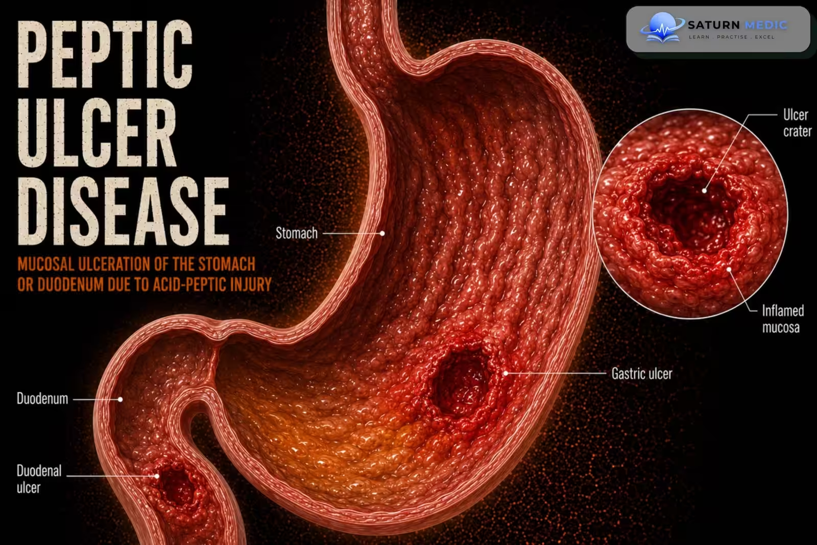
Peptic ulcer disease (PUD) is the presence of one or more ulcerative lesions in the stomach or
duodenum
Also called
ICD-10
Specialty
Onset
Reviewed
-
OverviewOverview
-
Etiology & Risk FactorsEtiology & Risk Factors
-
PathophysiologyPathophysiology
-
Clinical PresentationClinical Presentation
-
History TakingHistory Taking
-
Physical ExaminationPhysical Examination
-
InvestigationsInvestigations
-
DiagnosisDiagnosis
-
ManagementManagement
-
ComplicationsComplications
-
PrognosisPrognosis
-
Key Points / Clinical PearlsKey Points / Clinical Pearls
Overview
Peptic Ulcer Disease (PUD) is most commonly caused by Helicobacter pylori infection or prolonged NSAID
use. Other causes include hypersecretory states and stress. The typical symptom is epigastric pain,
although many patients are asymptomatic. Diagnosis is based on clinical evaluation, H. pylori
testing, and endoscopy when indicated. Treatment includes acid suppression, eradication of H.
pylori (if present), discontinuation of causative agents, and rarely surgery
Etiology & Risk Factors
Peptic ulcer disease is characterized by an open mucosal defect in the stomach or duodenum caused by an imbalance between damaging factors, such as gastric acid and pepsin, and protective mucosal mechanisms.
The two most common causes are:
- Helicobacter pylori infection
- Nonsteroidal anti-inflammatory drug use, including aspirin, ibuprofen, and naproxen
Other Causes
- Severe physiological stress in critically ill patients
- Zollinger–Ellison syndrome
- Corticosteroids, particularly when combined with NSAIDs
- Other medications that injure the gastric mucosa
- Crohn disease involving the stomach or duodenum
- Viral infections, such as cytomegalovirus, particularly in immunocompromised patients
- Gastric malignancy presenting as an ulcer
- Previous gastric surgery or radiation
Risk Factors
- Current or previous H. pylori infection
- Regular or high-dose NSAID use
- Older age
- Previous peptic ulcer or gastrointestinal bleeding
- Concurrent aspirin, anticoagulant, antiplatelet, or corticosteroid use
- Smoking
- Heavy alcohol consumption
- Chronic kidney disease
- Chronic liver disease
- Severe illness or ICU admission
- Family history of peptic ulcer disease
Pathophysiology
1. Breakdown of gastric or duodenal mucosal defenses.
2. Acid and pepsin damage the mucosa.
3. Ulcerative lesions develop in the stomach or duodenum.
4. Persistent injury may lead to ulcer complications if untreated.
Clinical Presentation
Common Symptoms
-
Burning or gnawing epigastric pain
-
Dyspepsia
-
Upper abdominal discomfort
-
Bloating
-
Belching
-
Nausea
-
Early satiety
-
Loss of appetite
-
Occasional vomiting
Duodenal Ulcer Presentation
-
Epigastric pain commonly occurs 2–5 hours after meals
-
Pain may improve temporarily after eating or taking antacids
-
Nocturnal pain is common
-
Symptoms may recur periodically
Gastric Ulcer Presentation
-
Epigastric pain may worsen shortly after eating
-
Early satiety may occur
-
Reduced food intake and weight loss may develop
-
Nausea and vomiting are more common than in duodenal ulcer
Features of Upper Gastrointestinal Bleeding
-
Hematemesis
-
Coffee-ground vomiting
-
Melena
-
Dizziness
-
Syncope
-
Fatigue due to anemia
-
Pallor
-
Tachycardia or hypotension in severe bleeding
Features of Perforation
-
Sudden, severe generalized abdominal pain
-
Rigid or board-like abdomen
-
Guarding and rebound tenderness
-
Absent bowel sounds
-
Signs of shock
Features of Gastric Outlet Obstruction
-
Persistent vomiting
-
Early satiety
-
Postprandial fullness
-
Abdominal distension
-
Weight loss
-
Dehydration
-
Succussion splash
Alarm Features
-
-
Unintentional weight loss
-
Iron-deficiency anemia
-
Persistent vomiting
-
Progressive dysphagia
-
Gastrointestinal bleeding
-
Palpable abdominal mass
-
New symptoms in an older patient
-
Family history of upper gastrointestinal malignancy
-
History Taking
-
Ask about:
• Epigastric pain
• Duration and pattern of symptoms
• NSAID use
• Previous history of peptic ulcers
• History suggestive of H. pylori infection
• Alarm symptoms or high-risk features
• Previous ulcer treatment
Physical Examination
Physical examination findings are variable and may be unremarkable.
Assess for abdominal tenderness and signs suggesting complications or alternative diagnoses.
Investigations
-
Initial Laboratory Tests
-
Complete blood count to detect anemia or infection
-
Electrolytes and renal function tests
-
Liver function tests
-
Serum amylase or lipase if pancreatitis is suspected
-
Coagulation profile if bleeding is present
-
Blood group and crossmatch in significant gastrointestinal bleeding
-
Stool occult blood testing when chronic blood loss is suspected
Helicobacter pylori Testing
Non-Invasive Tests
-
Urea breath test
-
Stool H. pylori antigen test
These tests identify active infection and may also be used to confirm eradication.
Invasive Tests During Endoscopy
-
Rapid urease test
-
Gastric biopsy with histology
-
Culture or molecular susceptibility testing in selected patients
Important Testing Note
PPIs may reduce the accuracy of urea breath and stool antigen tests. They are generally withheld for about two weeks when clinically safe, while antibiotics and bismuth are withheld for at least four weeks before testing.
Upper Gastrointestinal Endoscopy
Upper GI Endoscopy with Biopsy
Upper gastrointestinal endoscopy is the main investigation when direct visualization is required. It can:
-
Confirm the presence, location, size, and appearance of an ulcer
-
Identify active or recent bleeding
-
Permit endoscopic hemostasis
-
Obtain biopsies for H. pylori
-
Exclude gastric malignancy
Endoscopy and biopsy may be used to confirm an ulcer and determine its underlying cause.
Indications for Early Endoscopy
-
Gastrointestinal bleeding
-
Unintentional weight loss
-
Persistent vomiting
-
Progressive dysphagia
-
Iron-deficiency anemia
-
Palpable abdominal mass
-
Older patient with new symptoms
-
Failure of appropriate initial treatment
-
Suspected gastric ulcer or malignancy
Additional Investigations
-
CT abdomen and pelvis if perforation, penetration, obstruction, or another abdominal condition is suspected
-
Erect chest or abdominal X-ray may show free air in suspected perforation
-
Gastrin level if Zollinger–Ellison syndrome is suspected
-
Diagnosis
Diagnosis is confirmed by:
• EGD demonstrating the lesion
• Histopathological examination of biopsy tissue
Tumor staging is performed using:
• CT chest and abdomen
• PET scan
• Endoscopic ultrasound (EUS)
Related Topics
Management
1. Lifestyle Modifications
-
Stop NSAIDs if possible
-
Stop smoking
-
Avoid excessive alcohol
-
Avoid ulcer-triggering medications
2. Medical Therapy
Proton Pump Inhibitors (PPIs) – First-Line
-
Omeprazole
-
Pantoprazole
-
Esomeprazole
If H. pylori Positive
-
14-day eradication therapy
-
Continue PPI after treatment if indicated
3. Endoscopic Therapy
Indicated for:
-
Active upper GI bleeding
-
Recurrent bleeding
Treatment:
-
Endoscopic hemostasis (clips, thermal therapy, or injection)
4. Surgery
Indicated for:
-
Perforation
-
Uncontrolled bleeding
-
Gastric outlet obstruction
-
Refractory ulcers
5. Follow-Up
-
-
Confirm H. pylori eradication
-
Repeat endoscopy for gastric ulcers when indicated
-
Avoid NSAID recurrence
-
Complications
• Persistent or recurrent ulcers
• Need to exclude gastric malignancy in selected patients
Prognosis
- Most patients improve with appropriate acid suppression, eradication of H. pylori, and withdrawal of causative agents.
- Surgical intervention is rarely necessary. Some patients require surveillance if
symptoms persist or malignancy is suspected.
Key Points / Clinical Pearls
• Peptic Ulcer Disease (PUD) is an ulcer of the stomach or duodenum.
• The most common causes are H. pylori infection and NSAID use.
• Epigastric pain is the classic symptom, but many patients are asymptomatic.
• Patients <60 years without alarm features are often managed with H. pylori testing or empirical
PPI therapy.
• Patients ≥60 years or with high-risk features require EGD with biopsy.
• Treatment includes PPIs, H. pylori eradication (when indicated), and stopping NSAIDs.
• Acid suppression is continued for 4–8 weeks after eradication therapy.
• Surgery is rarely needed.
- National Center for Biotechnology Information (NIH). Peptic Ulcer Disease, StatPearls.
- Chey WD, Howden CW, Moss SF, et al. ACG Clinical Guideline: Treatment of Helicobacter pylori Infection. Am J Gastroenterol. 2024;119:1730-1753. DOI: 10.14309/ajg.0000000000002968.
- Laine L, Barkun AN, Saltzman JR, Martel M, Leontiadis GI. ACG Clinical Guideline: Upper Gastrointestinal and Ulcer Bleeding. Am J Gastroenterol. 2021;116:899-917. PMID: 33929377.
- Lanas A, Chan FKL. Peptic Ulcer Disease. Lancet. 2017;390:613-624. PMID: 28242110.
- MedlinePlus, National Library of Medicine (NIH). Peptic Ulcer: Health Topic.