

Clinical Subject Page
Intussusception
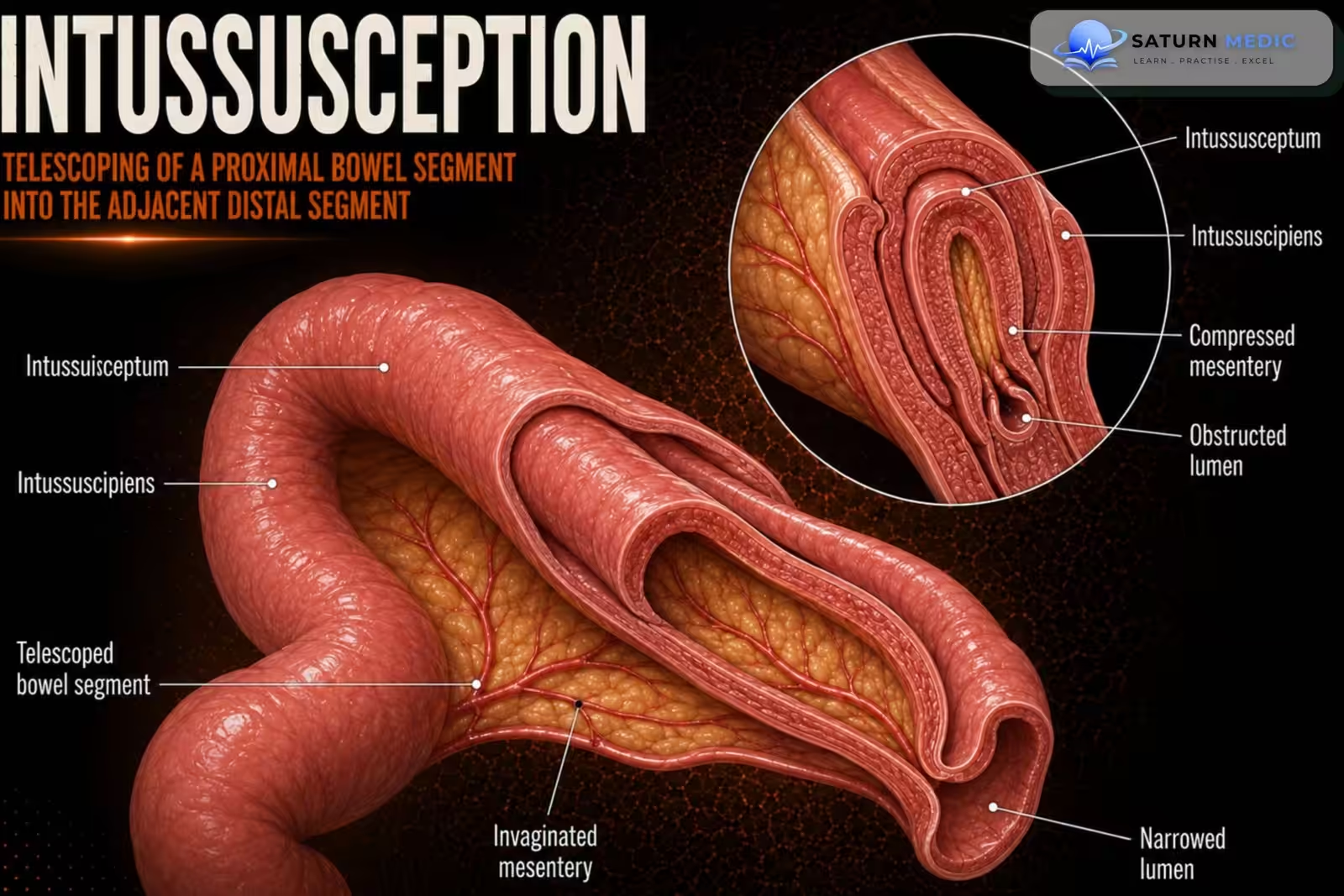
Intussusception is a condition in which one part of the intestine slides (telescopes) into the adjacent part of the intestine. This can block the bowel and reduce blood flow, leading to intestinal ischemia if not treated promptly.
Also called
ICD-10
Specialty
Onset
Reviewed
-
OverviewOverview
-
Etiology & Risk FactorsEtiology & Risk Factors
-
PathophysiologyPathophysiology
-
Clinical PresentationClinical Presentation
-
History TakingHistory Taking
-
Physical ExaminationPhysical Examination
-
InvestigationsInvestigations
-
DiagnosisDiagnosis
-
ManagementManagement
-
ComplicationsComplications
-
PrognosisPrognosis
-
Key Points / Clinical PearlsKey Points / Clinical Pearls
Overview
- Most common in infants and young children, and is usually idiopathic (no clear cause).
Typical symptoms include:
- Sudden, colicky (intermittent) abdominal pain
- Intermittent vomiting
- Child drawing the knees up to the chest
- Sometimes blood in the stool and a palpable abdominal mass
- Ultrasound is the preferred diagnostic test in children.
Etiology & Risk Factors
-Etiology
- Idiopathic (most common in children, ~90%)
- No identifiable cause.
- Often follows a viral illness (e.g., adenovirus), with enlarged Peyer patches acting as a temporary lead point.
- Pathological lead point
- Children (uncommon): Meckel diverticulum (most common), intestinal polyps, benign tumors, lymphoma, hematoma, hemangioma.
Adults (common): Usually caused by a neoplasm (tumor); other causes include infections, postoperative adhesions, Crohn disease, and congenital abnormalities.
-Risk Factors
- Age 3–12 months (highest risk in children)
- Recent viral illness
- Children > 5 years or recurrent intussusception (more likely to have a pathological lead point)
- Meckel diverticulum
- Cystic fibrosis
- IgA vasculitis (Henoch-Schönlein purpura)
- Intestinal tumors or polyps (especially in adults)
Pathophysiology
Lead point or enlarged Peyer patches → Intestinal telescoping (intussusception) → Bowel obstruction → Lymphatic obstruction → Venous congestion → Bowel wall edema → Reduced arterial blood flow → Intestinal ischemia → Necrosis (if untreated) → Perforation → Peritonitis
Clinical Presentation
-
Symptoms
- Sudden intermittent (colicky) abdominal pain
- Child draws knees to the chest during pain episodes
- Vomiting (may become bilious with bowel obstruction)
- Bloody or “currant jelly” stool (late sign, suggests bowel ischemia)
- Restlessness, irritability, or lethargy
Physical signs
- Abdominal tenderness
- Palpable sausage-shaped abdominal mass (usually in the right upper quadrant or epigastrium)
- Abnormal bowel sounds (high-pitched early, absent late)
History Taking
-
Ask about:
- When did the abdominal pain start?
- Is the pain intermittent (comes and goes) or constant?
- Does the child pull the knees to the chest during pain?
- Has the child been vomiting? Is it green (bilious)?
- Have you noticed blood or mucus in the stool?
- Has the child had diarrhea or constipation?
- Is the child irritable, restless, or unusually sleepy (lethargic)?
- Has the child had a recent viral illness (fever, cough, or cold)?
- Has this happened before?
- Does the child have any known bowel disease or previous abdominal surgery?
Physical Examination
- Assess vital signs (fever, tachycardia, signs of shock)
- Inspect the abdomen for distension
- Palpate for abdominal tenderness
- Feel for a sausage-shaped abdominal mass (usually in the right upper quadrant or epigastrium)
- Look for Dance sign (empty right lower quadrant; uncommon)
- Auscultate for high-pitched bowel sounds early or absent bowel sounds late
- Examine the stool for blood or “currant jelly” stool
Investigations
-
Laboratory Tests
- CBC – assess anemia or infection
- BMP (electrolytes) – assess dehydration and electrolyte imbalance
- Lactate – evaluate for bowel ischemia
Imaging
- Ultrasound abdomen (preferred in children)
- Target sign (concentric rings)
- Pseudokidney sign
- CT abdomen (preferred in adults)
- Target sign
- Detects lead point (e.g., tumor)
- Assesses for bowel ischemia
- Image-guided enema
- Used if diagnosis is uncertain and/or for reduction
- Contraindicated if bowel perforation is suspected
- Abdominal X-ray
- Not the initial test
- May show distended bowel loops if performed
Diagnosis
-
Diagnosis is based on clinical suspicion and confirmed with imaging.
- Children: Ultrasound abdomen (preferred)
- Target sign
- Pseudokidney sign
- Adults: CT abdomen (preferred)
- Target sign
- Identifies a pathological lead point and bowel ischemia
- Image-guided enema
- Used if diagnosis is uncertain and/or for reduction
- Do not use if bowel perforation is suspected
- Children: Ultrasound abdomen (preferred)
Related Topics
Management
-
Initial Management
- ABCDE assessment
- NPO (nothing by mouth)
- IV fluids for resuscitation
- Pain management
- Consider NG tube for gastric decompression
- Antibiotics if bowel ischemia or perforation is suspected
- Urgent surgical and/or radiology consultation
Definitive Treatment
- Clinically stable children:
- Image-guided pneumatic or hydrostatic enema reduction (first-line)
- Surgery if:
- Enema reduction fails
- Bowel perforation or intestinal ischemia is suspected
- Pathological lead point is present
- Most adult cases
After Successful Reduction
- Observe the child.
- Discharge if stable, asymptomatic, and tolerating oral fluids after observation.
- Admit if symptoms persist, reduction is unsuccessful, or risk factors for recurrence/complications are present.
Complications
- Bowel obstruction – blockage of intestinal contents.
- Intestinal ischemia – reduced blood supply leading to bowel injury.
- Bowel necrosis – death of bowel tissue if ischemia is prolonged.
- Gastrointestinal (bowel) perforation – rupture of the bowel wall.
- Peritonitis – inflammation/infection of the abdominal cavity after perforation.
- Recurrence – intussusception can recur after successful reduction.
Prognosis
- Intussusception is the telescoping of one part of the intestine into another.
- It is the most common cause of bowel obstruction in infants and young children.
- Most cases in children are idiopathic; adults usually have a pathological lead point (e.g., tumor).
- Classic symptoms: intermittent colicky abdominal pain, vomiting, and knees drawn to the chest.
- Currant jelly stool is a late sign and suggests bowel ischemia.
- Ultrasound is the diagnostic test of choice in children.
- CT abdomen is the diagnostic test of choice in adults.
- Pneumatic or hydrostatic enema is the first-line treatment in stable children.
- Surgery is required if enema reduction fails, perforation or ischemia is suspected, or in most adults.
Key Points / Clinical Pearls
- Vascular emergency with high mortality (50–70%).
- Most common cause: Superior mesenteric artery (SMA) embolism.
- Most important risk factor: Atrial fibrillation.
- Hallmark symptom: Severe abdominal pain out of proportion to the physical examination.
- Classic triad: Severe abdominal pain + bloody diarrhea + atrial fibrillation.
- CT angiography (CTA) is the test of choice.
- Do not delay CTA while waiting for laboratory results.
- Normal lactate does not exclude acute mesenteric ischemia early in the disease.
- Initial treatment: ABCDE, oxygen, IV fluids, NPO, NG tube, IV antibiotics, IV heparin (if appropriate), urgent surgical/vascular consultation.
- Peritonitis or hemodynamic instability = emergency laparotomy
- National Center for Biotechnology Information (NIH). Child Intussusception, StatPearls.
- Qiang H, Fu M, Feng S, et al. Repeated Ultrasound-Guided Hydrostatic Reduction for Pediatric Intussusception: Attempt Number, Interval, and Success. Eur J Med Res. 2025. PMID: 41291924.
- MedlinePlus, National Library of Medicine (NIH). Intussusception: Medical Encyclopedia.
- Determinants of Ultrasound-Guided Reduction Failure and Pathological Lead Points in Pediatric Intussusception. Pediatr Surg Int. 2025. PMC12909316.
- Gfroerer S, Fiegel H, Rolle U. Twenty Years' Experience for Reduction of Ileocolic Intussusceptions by Saline Enema Under Sonography Control. Pediatr Surg Int. 2016;32:679-682. PMID: 27154198.