

Clinical Subject Page
Meckel's Diverticulum
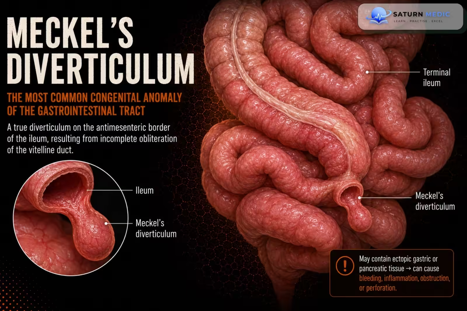
Meckel’s Diverticulum is the most common congenital abnormality of the gastrointestinal tract. It occurs because the vitelline (omphalomesenteric) duct fails to completely disappear during fetal development. It is a true diverticulum, meaning it contains all layers of the intestinal wall.
Also called
ICD-10
Specialty
Onset
Reviewed
-
OverviewOverview
-
Etiology & Risk FactorsEtiology & Risk Factors
-
PathophysiologyPathophysiology
-
Clinical PresentationClinical Presentation
-
History TakingHistory Taking
-
Physical ExaminationPhysical Examination
-
InvestigationsInvestigations
-
DiagnosisDiagnosis
-
ManagementManagement
-
ComplicationsComplications
-
PrognosisPrognosis
-
Key Points / Clinical PearlsKey Points / Clinical Pearls
Overview
- Usually found in the ileum, about 2 feet (60 cm) before the ileocecal valve.
- Often asymptomatic and discovered incidentally.
- May contain ectopic gastric mucosa, which can produce acid and cause ulceration and painless lower gastrointestinal bleeding, especially in children.
Etiology & Risk Factors
-Etiology
- Congenital condition caused by incomplete obliteration (failure of closure) of the vitelline (omphalomesenteric) duct during fetal development (normally closes by the 6th–7th week of gestation).
This results in a persistent true diverticulum of the ileum.
-Risk Factors
- Male sex (complications occur more often in males).
- Age < 2 years (symptoms are most common in young children, although it can present at any age).
- Presence of ectopic gastric mucosa (most common) or pancreatic tissue, which increases the risk of acid secretion, ulceration, and painless GI bleeding.
Pathophysiology
Failure of vitelline (omphalomesenteric) duct closure (6th–7th week of gestation) → Persistent true diverticulum in the ileum → May contain ectopic gastric (± pancreatic) mucosa → Acid secretion → Adjacent ileal ulceration → Painless lower GI bleeding
OR
Persistent Meckel’s diverticulum → Inflammation (diverticulitis) → Abdominal pain ± perforation
OR
Persistent Meckel’s diverticulum → Acts as a lead point or causes obstruction → Intussusception/bowel obstruction
Clinical Presentation
- Most patients are asymptomatic (found incidentally).
- Painless lower gastrointestinal bleeding (most common symptomatic presentation)
- Hematochezia (bright red blood per rectum)
- Melena (black, tarry stools)
- Abdominal pain (if diverticulitis develops; may mimic appendicitis)
- Bowel obstruction (vomiting, abdominal distension, constipation)
- Intussusception (may present with currant jelly stools)
- Signs of anemia (fatigue, pallor) due to chronic blood loss
- Perforation (rare) causing acute abdomen/peritonitis
History Taking
- Have you noticed blood in your stool? (bright red or black/tarry)
- Is the bleeding painful or painless?
- Do you have abdominal pain? Where is it located?
- Have you had vomiting or nausea?
- Do you have abdominal swelling or constipation?
- Have you had episodes of severe cramping abdominal pain?
- Have you noticed fatigue, weakness, or dizziness? (suggesting anemia)
- Have you had similar episodes before?
- Have you had previous abdominal surgery or known bowel problems?
- When did your symptoms start, and have they gotten worse?
Physical Examination
- General appearance: May be normal if asymptomatic
- Pallor (suggests anemia from GI bleeding)
- Vital signs: Tachycardia or hypotension if significant blood loss
- Abdominal tenderness (especially if diverticulitis is present)
- Abdominal distension (suggests bowel obstruction)
- Signs of peritonitis (guarding, rigidity, rebound tenderness) if perforation occurs
- Rectal examination: Bright red blood or melena may be present
Investigations
Laboratory tests
- Complete blood count (CBC): Check for anemia from blood loss.
- Investigations are guided by the presentation (e.g., GI bleeding or acute abdomen).
Imaging
- Meckel scan (Technetium-99m pertechnetate scan) – Investigation of choice, especially in children; detects ectopic gastric mucosa.
- Abdominal ultrasound (US): Assesses complications such as intussusception or diverticulitis.
- CT abdomen with IV contrast: Evaluates diverticulitis, bowel obstruction, perforation, and helps exclude other diagnoses.
- CT angiography: Used to localize the source of active GI bleeding.
Other tests
- Endoscopy (capsule or double-balloon enteroscopy): May help visualize the diverticulum if the diagnosis is uncertain.
- Diagnostic laparoscopy: Consider if the diagnosis remains unclear.
Diagnosis
- Diagnosis is based on clinical suspicion and confirmed with imaging, especially in patients with painless lower GI bleeding.
- Diagnostic approach:
- Suspect Meckel’s diverticulum in a child with painless lower GI bleeding or unexplained acute abdomen.
- Meckel scan (Technetium-99m pertechnetate scan) is the test of choice, particularly in children.
- Ultrasound or CT abdomen helps identify complications (diverticulitis, intussusception, bowel obstruction, perforation) and exclude other causes.
- CT angiography is used if there is active GI bleeding.
- Capsule endoscopy, double-balloon enteroscopy, or diagnostic laparoscopy may be used if the diagnosis remains uncertain.
Related Topics
Management
-Initial management
- Stabilize the patient (ABCDE) if unstable.
- Treat GI bleeding or acute abdomen as needed.
- Consult surgery if Meckel’s diverticulum is confirmed or strongly suspected.
-Definitive treatment
- Symptomatic Meckel’s diverticulum → Surgical resection
- Diverticulectomy
- Segmental bowel resection (if required)
- Asymptomatic Meckel’s diverticulum
- Found on imaging: No treatment needed.
- Found incidentally during surgery:
- Children: Usually resect.
- Adults: Individualized decision based on risks and benefits.
Complications
- GI hemorrhage (most common complication) → painless lower GI bleeding
- Meckel diverticulitis → mimics acute appendicitis
- Bowel obstruction
- Intussusception
- Volvulus
- Littré hernia
- Bowel perforation → peritonitis or intra-abdominal abscess
- Anemia due to chronic blood loss
- Neoplasia (rare)
Prognosis
- Excellent prognosis if diagnosed early and treated appropriately.
- Most patients recover completely after surgical resection.
- Asymptomatic Meckel’s diverticulum usually remains harmless and often requires no treatment if found only on imaging.
- Prognosis becomes less favorable if complications (e.g., bleeding, bowel obstruction, diverticulitis, or perforation) are not recognized and treated promptly.
Key Points / Clinical Pearls
- Most common congenital abnormality of the GI tract.
- Caused by failure of the vitelline (omphalomesenteric) duct to close.
- It is a true diverticulum and is usually located 2 feet from the ileocecal valve.
- May contain ectopic gastric mucosa, causing acid secretion → ileal ulceration → painless lower GI bleeding.
- Most patients are asymptomatic.
- Most common symptomatic presentation: Painless lower GI bleeding, especially in children < 2 years.
- Other complications include diverticulitis, bowel obstruction, intussusception, perforation, and anemia.
- Meckel scan (Technetium-99m) is the investigation of choice, especially in children.
- National Center for Biotechnology Information (NIH). Small Bowel Neoplasms, StatPearls.
- Boudreaux JP, Klimstra DS, Hassan MM, et al; North American Neuroendocrine Tumor Society (NANETS). The NANETS Consensus Guideline for the Diagnosis and Management of Neuroendocrine Tumors: Well-Differentiated Neuroendocrine Tumors of the Jejunum, Ileum, Appendix, and Cecum. Pancreas. 2010;39:753-766. PMID: 20664473.
- Howe JR, Cardona K, Fraker DL, et al. The Surgical Management of Small Bowel Neuroendocrine Tumors: Consensus Guidelines of the North American Neuroendocrine Tumor Society (NANETS). Pancreas. 2017;46:715-731. PMC5502737.
- Diagnosis and Management of Small Bowel Neuroendocrine Tumors: A State-of-the-Art Review. PMC9516545.
- MedlinePlus, National Library of Medicine (NIH). Carcinoid Syndrome: Medical Encyclopedia.