

Clinical Subject Page
Colorectal Cancer
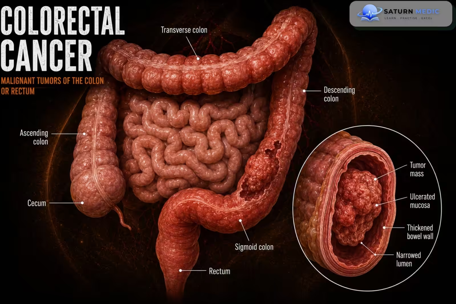
Colorectal cancer (CRC) is a malignant tumor arising from the colon or rectum, most commonly developing from adenomatous polyps through the adenoma–carcinoma sequence. It is one of the most common cancers worldwide and is highly treatable when detected early
Also called
ICD-10
Specialty
Onset
Reviewed
-
OverviewOverview
-
Etiology & Risk FactorsEtiology & Risk Factors
-
PathophysiologyPathophysiology
-
Clinical PresentationClinical Presentation
-
History TakingHistory Taking
-
Physical ExaminationPhysical Examination
-
InvestigationsInvestigations
-
DiagnosisDiagnosis
-
ManagementManagement
-
ComplicationsComplications
-
PrognosisPrognosis
-
Key Points / Clinical PearlsKey Points / Clinical Pearls
Overview
Colorectal cancer develops through the progressive accumulation of genetic mutations that transform normal colonic mucosa into adenomatous polyps and eventually invasive carcinoma. Most tumors are adenocarcinomas. Patients may present with rectal bleeding, altered bowel habits, iron-deficiency anemia, abdominal pain, or bowel obstruction. Diagnosis is confirmed by colonoscopy with biopsy, and staging is performed using CT imaging. Treatment depends on the stage and typically includes surgery, chemotherapy, radiotherapy (for rectal cancer), and targeted or immunotherapy in selected patients.
Etiology & Risk Factors
Aetiology
Most colorectal cancers arise from adenomatous polyps through the adenoma–carcinoma sequence involving progressive genetic mutations.
Risk Factors
Non-Modifiable
Age >50 years
Family history of colorectal cancer
Familial adenomatous polyposis (FAP)
Lynch syndrome (HNPCC)
Personal history of colorectal cancer
Personal history of adenomatous polyps
Long-standing ulcerative colitis
Long-standing Crohn’s colitis
Modifiable
Diet high in red or processed meat
Low-fiber diet
Obesity
Physical inactivity
Smoking
Heavy alcohol consumption
Type 2 diabetes
Pathophysiology
Most colorectal cancers develop through the adenoma–carcinoma sequence. Progressive genetic mutations, including alterations in APC, KRAS, and TP53, transform normal colonic epithelium into adenomatous polyps, which gradually progress to invasive adenocarcinoma. As the tumor enlarges, it invades the bowel wall, spreads to regional lymph nodes, and may metastasize to distant organs, particularly the liver and lungs
Clinical Presentation
Symptoms
Rectal bleeding
Occult gastrointestinal bleeding
Iron-deficiency anemia
Change in bowel habits
Constipation
Diarrhea
Narrow stools
Abdominal pain
Bloating
Tenesmus (rectal cancer)
Weight loss
Fatigue
Signs
Pallor
Abdominal mass
Rectal mass
Hepatomegaly (liver metastasis)
Ascites (advanced disease)
Intestinal obstruction
Cachexia
History Taking
Ask about:
- Rectal bleeding
- Stool color
- Change in bowel habits
- Duration of symptoms
- Abdominal pain
- Weight loss
- Fatigue
- Symptoms of anemia
- Family history of colorectal cancer
- Personal history of polyps
- Inflammatory bowel disease
- Previous colonoscopy
Physical Examination
General Examination
Look for:
Pallor
Weight loss
Cachexia
Lymphadenopathy
Abdominal Examination
Assess for:
Abdominal tenderness
Distension
Palpable abdominal mass
Hepatomegaly
Ascites
Digital Rectal Examination (DRE)
Assess for:
Rectal mass
Blood on the glove
Rectal tenderness
Anal pathology
Other Examination
Look for:
Virchow’s node
Signs of liver metastases
Signs of bowel obstruction
Investigations
-Laboratory Tests
Complete blood count
Iron studies
Liver function tests
Renal function
Electrolytes
CEA (Carcinoembryonic antigen)
-Colonoscopy
Gold standard investigation
Allows:
Direct visualization
Biopsy
Detection of synchronous lesions
Polypectomy when appropriate
-Histopathology
Confirms:
Adenocarcinoma
Tumor grade
Histological subtype
-Imaging
CT Chest, Abdomen and Pelvis
Used for:
TNM staging
Detecting metastases
Surgical planning
MRI Pelvis
Preferred for:
Rectal cancer staging
Circumferential resection margin assessment
Endorectal Ultrasound
Useful for:
Early rectal cancer staging
PET-CT
Selected cases:
Suspected metastatic disease
Recurrent disease
Diagnosis
Early Disease (Stage I)
Surgical resection
Endoscopic resection for selected very early lesions
Localized Disease (Stage II–III Colon Cancer)
Curative surgical resection
Adjuvant chemotherapy for selected Stage II and most Stage III patients
Rectal Cancer
May require:
Neoadjuvant chemoradiotherapy or total neoadjuvant therapy
Surgical resection
Adjuvant chemotherapy when indicated
Metastatic Disease
Treatment may include:
Systemic chemotherapy
Targeted therapy
Immunotherapy (MSI-H/dMMR tumors)
Liver or lung metastasectomy in selected patients
Palliative surgery when necessary
Supportive Care
Nutritional support
Pain management
Stoma care
Psychological support
Palliative care for advanced disease
Related Topics
Management
1. Control Gastroesophageal Reflux
Give a proton pump inhibitor (PPI), usually once daily.
Increase to twice daily when reflux symptoms remain uncontrolled or during endoscopic treatment when indicated.
Encourage:
Weight reduction when overweight
Smoking cessation
Avoiding late meals
Elevating the head of the bed for nocturnal reflux
Avoiding individual reflux triggers
Antireflux surgery is considered for persistent reflux despite optimized medical therapy, but it is not recommended solely to prevent cancer.
2. Endoscopic Surveillance
Non-dysplastic Barrett’s Esophagus
No immediate ablation is required.
Continue PPI therapy.
Perform periodic surveillance upper endoscopy with systematic biopsies.
Surveillance intervals are usually based on the length of the Barrett’s segment:
<3 cm: approximately every 5 years
≥3 cm: approximately every 3 years
Indefinite for Dysplasia
Confirm the diagnosis with an expert gastrointestinal pathologist.
Optimize acid suppression, often with twice-daily PPI.
Repeat endoscopy with biopsies after approximately 6 months.
Persistent indefinite dysplasia requires closer surveillance.
3. Low-Grade Dysplasia
Confirm the diagnosis by a second expert gastrointestinal pathologist.
Endoscopic eradication therapy is generally preferred.
Endoscopic surveillance remains an alternative in selected patients after discussing risks and benefits.
Common eradication treatment:
Endoscopic resection of visible lesions
Radiofrequency ablation of the remaining Barrett’s mucosa
4. High-Grade Dysplasia
Refer to an experienced specialist or high-volume center.
Perform careful endoscopic assessment.
Remove visible or nodular lesions using:
Endoscopic mucosal resection
Endoscopic submucosal dissection in selected cases
Ablate the remaining Barrett’s epithelium, commonly with radiofrequency ablation.
High-grade dysplasia should generally receive endoscopic eradication therapy because of its substantial risk of progression to adenocarcinoma.
5. Early Esophageal Adenocarcinoma
For superficial cancer limited to the mucosa:
Endoscopic resection of the cancer
Ablation of the remaining Barrett’s mucosa
Close surveillance afterward
Esophagectomy may be required when there is:
Deep submucosal invasion
Lymphovascular invasion
Poor differentiation
Positive deep resection margins
Suspected lymph-node involvement
Disease unsuitable for endoscopic treatment
6. Surveillance After Eradication
Even after complete eradication of intestinal metaplasia:
Continue PPI therapy.
Perform scheduled surveillance endoscopy.
Carefully inspect the gastroesophageal junction and previous Barrett’s segment.
Biopsy suspicious areas because recurrence can occur.
Complications
- Bowel obstruction
- Bowel perforation
- Gastrointestinal bleeding
- Iron-deficiency anemia
- Liver metastases
- Lung metastases
- Peritoneal metastases
- Local recurrence
- Cancer cachexia
- Venous thromboembolism
Prognosis
- The prognosis depends primarily on the TNM stage at diagnosis. Early-stage disease has an excellent prognosis following complete surgical resection, whereas metastatic disease has a significantly poorer outcome. Regular surveillance after treatment improves the detection of recurrence and new primary tumors.
Key Points / Clinical Pearls
- Colorectal cancer usually develops from adenomatous polyps.
- Adenocarcinoma accounts for over 90% of cases.
- Rectal bleeding and altered bowel habits are common presenting symptoms.
- Right-sided tumors often cause iron-deficiency anemia.
- Colonoscopy with biopsy is the diagnostic gold standard.
- CT is used for staging, while MRI is preferred for rectal cancer staging.
- Surgical resection is the main curative treatment.
- Chemotherapy, radiotherapy, targeted therapy, and immunotherapy are used according to disease stage.
- Early detection through screening significantly improves survival.
- Menon G, Cagir B. National Center for Biotechnology Information (NIH). Colon Cancer, StatPearls.
- US Preventive Services Task Force. Screening for Colorectal Cancer: US Preventive Services Task Force Recommendation Statement. JAMA. 2021;325:1965-1977. USPSTF Recommendation.
- National Cancer Institute (NIH). Colon Cancer Treatment (PDQ), Health Professional Version.
- Kumar R, Lewis CR. National Center for Biotechnology Information (NIH). Colon Cancer Screening, StatPearls.
- National Center for Biotechnology Information (NIH). Rectal Cancer, StatPearls.