

Clinical Subject Page
Ischemic colitis
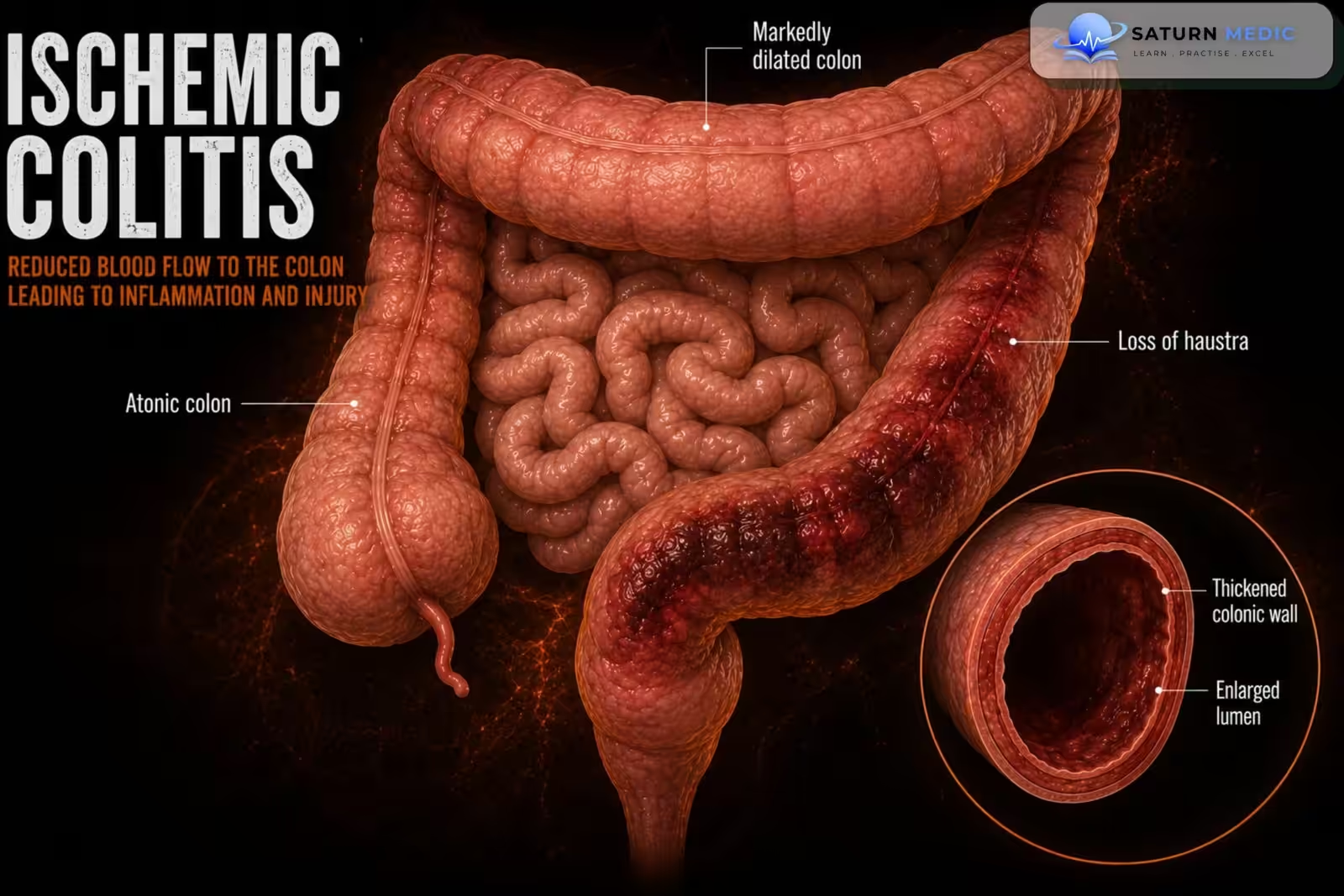
Ischemic colitis is acute inflammation and injury of the colon caused by reduced blood flow, resulting in mucosal ischemia. It is the most common form of intestinal ischemia and usually affects the left colon, particularly the splenic flexure and sigmoid colon
Also called
ICD-10
Specialty
Onset
Reviewed
-
OverviewOverview
-
Etiology & Risk FactorsEtiology & Risk Factors
-
PathophysiologyPathophysiology
-
Clinical PresentationClinical Presentation
-
History TakingHistory Taking
-
Physical ExaminationPhysical Examination
-
InvestigationsInvestigations
-
DiagnosisDiagnosis
-
ManagementManagement
-
ComplicationsComplications
-
PrognosisPrognosis
-
Key Points / Clinical PearlsKey Points / Clinical Pearls
Overview
Ischemic colitis occurs when blood flow to the colon is reduced, leading to ischemia and inflammation of the bowel wall. Most cases are transient and resolve with supportive treatment, but severe ischemia can progress to transmural necrosis, perforation, peritonitis, and sepsis. Diagnosis is based on clinical presentation, CT imaging, and colonoscopy with biopsy when appropriate
Etiology & Risk Factors
Etiology
Reduced colonic blood flow due to:
-
Non-occlusive hypoperfusion (most common)
-
Arterial thrombosis
-
Arterial embolism
-
Mesenteric venous thrombosis (rare cause)
-
Mechanical obstruction affecting blood supply
-
Vasculitis
Risk Factors
-
Age >60 years
-
Atherosclerosis
-
Heart failure
-
Hypotension or shock
-
Dehydration
-
Diabetes mellitus
-
Hypertension
-
Chronic kidney disease
-
Atrial fibrillation
-
Hypercoagulable disorders
-
Previous vascular disease
-
Recent major surgery
-
Cocaine or methamphetamine use
-
Estrogen-containing medications
-
Smoking
Pathophysiology
Reduced colonic blood flow causes inadequate oxygen delivery to the bowel wall, leading initially to mucosal ischemia and inflammation. If blood flow is restored promptly, the injury is often reversible. Persistent ischemia progresses to full-thickness bowel necrosis, bacterial translocation, perforation, peritonitis, sepsis, and multiorgan failure
Clinical Presentation
Symptoms
-
Sudden crampy abdominal pain
-
Left lower-quadrant pain
-
Urgency to defecate
-
Bloody diarrhea
-
Hematochezia
-
Mild fever
-
Nausea
-
Vomiting
Signs
-
Left-sided abdominal tenderness
-
Mild abdominal distension
-
Rectal bleeding
-
Tachycardia
-
Fever
-
Guarding or rebound tenderness (advanced disease)
-
Hypotension in severe cases
-
Sudden crampy abdominal pain
-
Left lower-quadrant pain
-
Urgency to defecate
-
Bloody diarrhea
-
Hematochezia
-
Mild fever
-
Nausea
-
Vomiting
Signs
-
-
Left-sided abdominal tenderness
-
Mild abdominal distension
-
Rectal bleeding
-
Tachycardia
-
Fever
-
Guarding or rebound tenderness (advanced disease)
-
Hypotension in severe cases
-
History Taking
Ask about:
- Onset of abdominal pain
- Location of pain
- Bloody stools
- Diarrhea
- Previous similar episodes
- Recent hypotension or dehydration
- Cardiac disease
- Atrial fibrillation
- Vascular disease
- Recent surgery
- Medication history
Physical Examination
General Examination
Look for:
-
Fever
-
Tachycardia
-
Hypotension
-
Dehydration
-
Pallor
-
Signs of shock
Abdominal Examination
Assess for:
-
Left-sided tenderness
-
Mild distension
-
Guarding
-
Rebound tenderness
-
Absent bowel sounds (advanced ischemia)
-
Peritoneal signs
Rectal Examination
Assess for:
-
Gross blood
-
Occult blood
-
Rectal tenderness
Investigations
Laboratory Tests
-
Complete blood count
-
C-reactive protein
-
ESR
-
Renal function and electrolytes
-
Liver function tests
-
Serum lactate
-
Arterial blood gas
-
Coagulation profile
-
Blood cultures if septic
Possible findings:
-
Leukocytosis
-
Elevated CRP
-
Elevated lactate (severe ischemia)
-
Metabolic acidosis
-
Anemia if significant bleeding
CT Abdomen and Pelvis with IV Contrast
First-line imaging
Typical findings:
-
Segmental colonic wall thickening
-
Thumbprinting
-
Pericolic fat stranding
-
Bowel-wall edema
-
Reduced bowel enhancement
-
Pneumatosis intestinalis (advanced disease)
-
Portal venous gas (severe ischemia)
Colonoscopy
Usually performed within 24–48 hours in stable patients.
Findings include:
-
Pale or edematous mucosa
-
Petechiae
-
Longitudinal ulcerations
-
Cyanotic mucosa
-
Friability
-
Sharp transition between normal and ischemic bowel
Biopsy confirms ischemic injury and excludes inflammatory bowel disease or malignancy.
CT Angiography
Usually reserved for suspected acute mesenteric ischemia or when major arterial occlusion is suspected.
Diagnosis
Diagnosis is based on:
- Typical clinical presentation
- CT findings
- Colonoscopy with biopsy
- Exclusion of infectious and inflammatory causes
- Identification of underlying vascular risk factors
Related Topics
Management
Mild to Moderate Disease
-
Hospital admission
-
Bowel rest
-
Intravenous fluids
-
Correction of electrolyte abnormalities
-
Oxygen if needed
-
Treat underlying cause
-
Stop vasoconstrictive medications when appropriate
-
Close clinical observation
Antibiotics
Broad-spectrum intravenous antibiotics are commonly recommended for:
-
Moderate disease
-
Severe disease
-
Suspected transmural ischemia
-
Peritonitis
-
Immunocompromised patients
Severe Disease
-
Intensive care monitoring
-
Aggressive fluid resuscitation
-
Broad-spectrum IV antibiotics
-
Serial abdominal examinations
-
Repeat imaging if deterioration occurs
Surgery
Indications include:
-
Peritonitis
-
Colonic perforation
-
Bowel gangrene
-
Persistent gastrointestinal bleeding
-
Clinical deterioration
-
Fulminant colitis
-
Transmural necrosis
-
Failure of conservative treatment
Procedures may include:
-
Segmental colectomy
-
Subtotal colectomy
-
Temporary stoma depending on bowel viability
Complications
- Colonic necrosis
- Bowel perforation
- Peritonitis
- Sepsis
- Septic shock
- Colonic stricture
- Chronic ischemic colitis
- Recurrent ischemia
- Death
Prognosis
Most patients with mild ischemic colitis recover completely within a few days with conservative management. Prognosis is poor in patients with transmural infarction, right-sided colonic involvement, delayed diagnosis, septic shock, or those requiring emergency surgery
Key Points / Clinical Pearls
- Ischemic colitis is the most common form of intestinal ischemia.
- It usually affects the splenic flexure and sigmoid colon.
- Sudden left-sided abdominal pain followed by bloody diarrhea is the classic presentation.
- CT abdomen with IV contrast is the preferred initial investigation.
- Colonoscopy confirms the diagnosis in stable patients.
- Most patients recover with bowel rest, IV fluids, and supportive care.
- Broad-spectrum antibiotics are used in moderate-to-severe disease.
- Perforation, gangrene, or peritonitis requires emergency surgery.
- Right-sided ischemic colitis is associated with a worse prognosis.
- Amini A, Nagalli S. National Center for Biotechnology Information (NIH). Bowel Ischemia, StatPearls.
- Brandt LJ, Feuerstadt P, Longstreth GF, Boley SJ; American College of Gastroenterology. ACG Clinical Guideline: Epidemiology, Risk Factors, Patterns of Presentation, Diagnosis, and Management of Colon Ischemia (CI). Am J Gastroenterol. 2015;110:18-44. PMID: 25559486.
- Yadav S, Dave M, Edakkanambeth Varayil J, et al. A Population-Based Study of Incidence, Risk Factors, Clinical Spectrum, and Outcomes of Ischemic Colitis. Clin Gastroenterol Hepatol. 2015;13:731-738. PMID: 25130937.
- MedlinePlus, National Library of Medicine (NIH). Ischemic Colitis: Medical Encyclopedia.
- Azer SA, Sun Y. National Center for Biotechnology Information (NIH). Colitis, StatPearls.