

Clinical Subject Page
Pseudomembranous Colitis
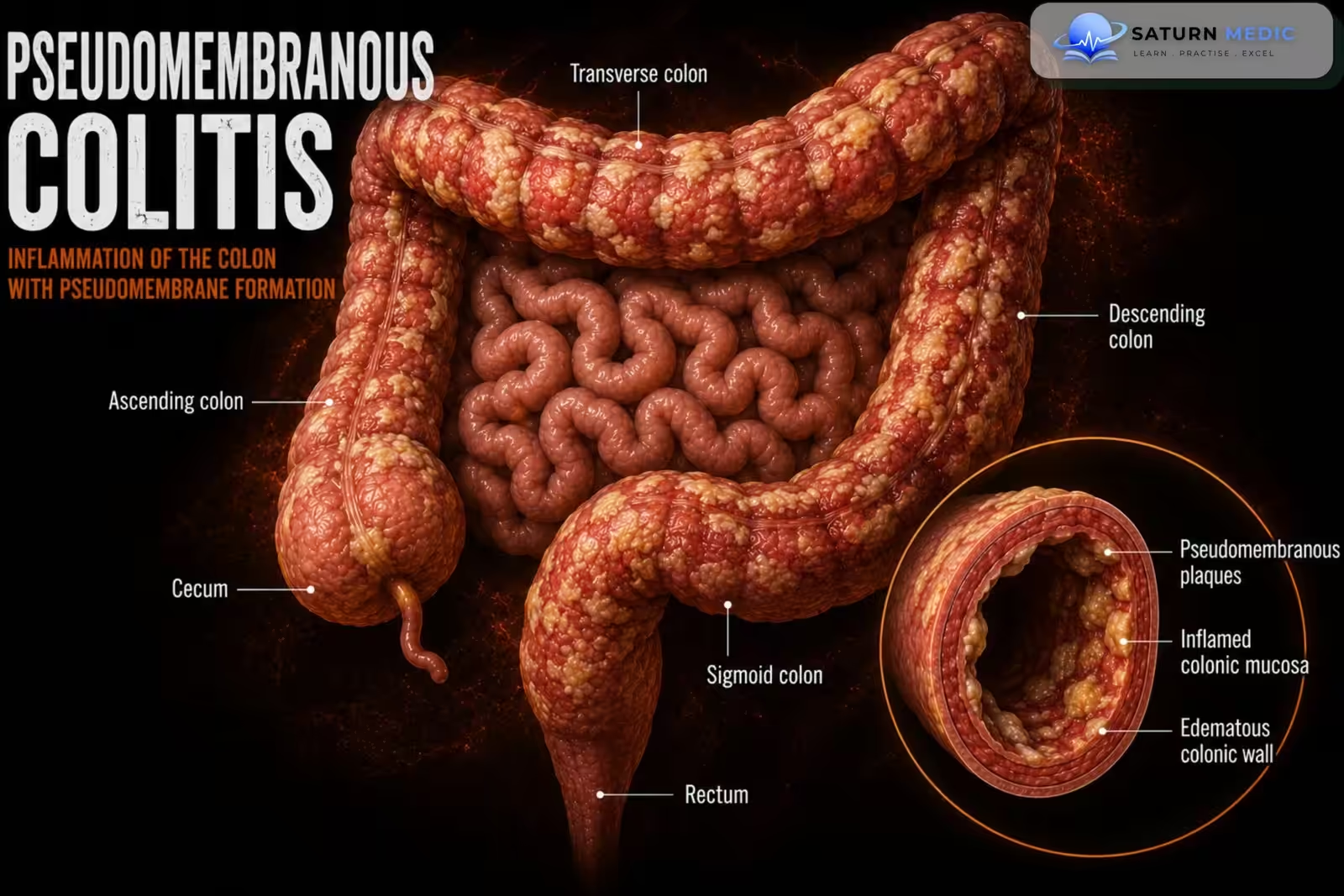
Pseudomembranous colitis is an inflammatory disease of the colon most commonly caused by toxin-producing Clostridioides difficile. It usually develops after antibiotic therapy disrupts the normal intestinal microbiota, allowing C. difficile to multiply and release toxins that cause watery diarrhea and colonic inflammation
Also called
ICD-10
Specialty
Onset
Reviewed
-
OverviewOverview
-
Etiology & Risk FactorsEtiology & Risk Factors
-
PathophysiologyPathophysiology
-
Clinical PresentationClinical Presentation
-
History TakingHistory Taking
-
Physical ExaminationPhysical Examination
-
InvestigationsInvestigations
-
DiagnosisDiagnosis
-
ManagementManagement
-
ComplicationsComplications
-
PrognosisPrognosis
-
Key Points / Clinical PearlsKey Points / Clinical Pearls
Overview
Ulcerative colitis causes relapsing and remitting inflammation limited primarily to the inner lining of the colon. The disease typically starts in the rectum and spreads proximally in a continuous pattern. Patients may experience periods of active symptoms followed by remission. Diagnosis is based on clinical findings, stool testing, colonoscopy, and biopsy. Treatment aims to induce and maintain remission, heal the mucosa, prevent complications, and improve quality of life
Etiology & Risk Factors
Etiology
Pseudomembranous colitis is caused mainly by toxin-producing Clostridioides difficile.
Important bacterial toxins include:
Toxin A: enterotoxin
Toxin B: cytotoxin
Binary toxin in some highly virulent strains
Commonly Associated Antibiotics
Clindamycin
Cephalosporins
Fluoroquinolones
Broad-spectrum penicillins
Carbapenems
Almost any antibiotic can cause C. difficile infection.
Risk Factors
Recent or prolonged antibiotic use
Recent hospitalization
Residence in a nursing or long-term care facility
Advanced age
Previous C. difficile infection
Immunosuppression
Severe underlying illness
Gastrointestinal surgery
Prolonged healthcare exposure
Inflammatory bowel disease
Acid-suppressive medication, particularly when unnecessary
Pathophysiology
Antibiotic exposure disrupts the normal colonic microbiota, reducing resistance to colonization by C. difficile. Ingested spores survive gastric acid, reach the colon, and germinate into toxin-producing bacteria. Toxins damage colonic epithelial cells, disrupt tight junctions, and trigger intense neutrophilic inflammation. Fluid and electrolytes enter the intestinal lumen, causing watery diarrhea. Necrotic epithelial cells, mucus, fibrin, and inflammatory cells accumulate on the mucosal surface and form characteristic pseudomembranes. Severe inflammation may progress to ileus, toxic megacolon, perforation, sepsis, and shock
Clinical Presentation
Common Symptoms
Frequent watery diarrhea
Lower abdominal cramps
Abdominal tenderness
Fever
Nausea
Loss of appetite
Malaise
Dehydration
Severe or Fulminant Disease
Marked abdominal pain
Abdominal distension
High fever
Tachycardia
Hypotension
Reduced urine output
Ileus
Altered mental status
Toxic megacolon
Peritonitis
Septic shock
History Taking
Ask about:
- Onset and frequency of diarrhea
- Stool consistency
- Presence of blood or mucus
- Abdominal pain or distension
- Fever or chills
- Nausea or vomiting
- Recent antibiotic use
- Type and duration of antibiotics
- Recent hospitalization
- Nursing-home residence
- Previous C. difficile infection
- Immunosuppressive medication
- Inflammatory bowel disease
Physical Examination
General Examination
Assess for:
Fever
Tachycardia
Hypotension
Dehydration
Pallor
Confusion
Sepsis or shock
Abdominal Examination
Look for:
Abdominal distension
Diffuse or lower abdominal tenderness
Tympany
Reduced bowel sounds in ileus
Guarding
Rebound tenderness
Signs of peritonitis
Investigations
Stool Testing
Test patients who have new, unexplained, clinically significant diarrhea, usually three or more unformed stools within 24 hours.
Available tests include:
Glutamate dehydrogenase antigen test
Toxin A and B enzyme immunoassay
Nucleic acid amplification test or PCR
Multistep testing algorithms combining antigen, toxin, and molecular tests
Only unformed stool should generally be tested. Testing asymptomatic patients may detect colonization rather than active infection.
Laboratory Tests
Complete blood count
C-reactive protein
Renal function and electrolytes
Serum albumin
Serum lactate
Liver function tests
Blood cultures if septic
Arterial or venous blood gas in severe disease
Possible findings include:
Leukocytosis
Acute kidney injury
Hypokalemia
Hypoalbuminemia
Elevated inflammatory markers
Metabolic acidosis
Elevated lactate
Imaging
Abdominal Radiograph
May show:
Colonic dilatation
Ileus
Toxic megacolon
Free air if perforation has occurred
CT Abdomen and Pelvis
Consider in severe or complicated disease.
Possible findings include:
Colonic wall thickening
Pericolic fat stranding
Mucosal edema
Ascites
Colonic dilatation
Toxic megacolon
Perforation
Endoscopy
Colonoscopy is not routinely required.
It may be considered when:
Stool tests are inconclusive
Another diagnosis is suspected
Rapid confirmation is essential
Typical findings include raised yellow-white pseudomembranes over inflamed colonic mucosa.
Endoscopy should be avoided in toxic megacolon or suspected perforation.
Diagnosis
Diagnosis
Diagnosis requires:
Compatible symptoms, particularly new-onset diarrhea
Positive testing for toxigenic C. difficile or its toxins
Exclusion of alternative causes when necessary
Severity Classification
Non-Severe Infection
Leukocytosis not exceeding approximately 15,000 cells/mm³
Serum creatinine below 1.5 mg/dL
Severe Infection
White blood cell count above 15,000 cells/mm³, or
Serum creatinine of at least 1.5 mg/dL
Fulminant Infection
One or more of:
Hypotension
Shock
Ileus
Toxic megacolon
Related Topics
Management
General Measures
Stop the precipitating antibiotic whenever clinically possible
Stop unnecessary proton-pump inhibitors
Correct dehydration and electrolyte abnormalities
Avoid antimotility drugs such as loperamide
Monitor vital signs and urine output
Assess for ileus, megacolon, or perforation
Use contact precautions
Seek early surgical advice in fulminant disease
Initial Non-Fulminant Infection
Preferred treatment:
Oral fidaxomicin
Acceptable alternative:
Oral vancomycin
IDSA and SHEA suggest fidaxomicin rather than a standard course of vancomycin for an initial episode when available, while recognizing vancomycin as an acceptable alternative.
Metronidazole is generally not preferred when fidaxomicin or vancomycin is available.
Fulminant Infection
Management generally includes:
High-dose oral or nasogastric vancomycin
Intravenous metronidazole
Rectal vancomycin when severe ileus prevents adequate colonic drug delivery
Aggressive intravenous fluid resuscitation
Intensive monitoring
Immediate surgical consultation
Recurrent Infection
Options depend on previous treatment and recurrence history:
Fidaxomicin
Vancomycin taper-and-pulse regimen
Vancomycin followed by rifaximin in selected patients
Bezlotoxumab for selected patients at high risk of further recurrence
Fecal microbiota-based therapy after multiple recurrences despite appropriate antibiotics
IDSA and SHEA recommend considering fidaxomicin for recurrent infection and bezlotoxumab as an adjunct in selected patients with recurrence risk.
Surgical Management
Urgent surgery may be required for:
Toxic megacolon
Colonic perforation
Peritonitis
Bowel necrosis
Progressive shock
Worsening organ failure
Failure of maximal medical treatment
Procedures may include:
Subtotal colectomy with end ileostomy
Diverting loop ileostomy with colonic lavage in selected patients
Complications
- Severe dehydration
- Electrolyte abnormalities
- Acute kidney injury
- Hypoalbuminemia
- Ileus
- Toxic megacolon
- Colonic perforation
- Peritonitis
- Sepsis
- Septic shock
- Recurrent infection
- Multiorgan failure
- Death
Prognosis
Most patients improve with appropriate antibiotic therapy and withdrawal of the precipitating antibiotic. Prognosis is worse in older adults, immunocompromised patients, those with severe leukocytosis or renal failure, and patients who develop ileus, toxic megacolon, perforation, or shock. Recurrence is common and becomes increasingly likely after each repeated episode.
Key Points / Clinical Pearls
- Pseudomembranous colitis is usually caused by toxin-producing C. difficile.
- Recent antibiotic exposure is the major risk factor.
- Watery diarrhea, abdominal pain, fever, and leukocytosis are typical.
- Test only patients with clinically significant unformed stools.
- A positive molecular test alone may represent colonization if symptoms are absent.
- Fidaxomicin is generally preferred for an initial non-fulminant episode when available.
- Oral vancomycin remains an effective alternative.
- Fulminant infection requires high-dose oral vancomycin, IV metronidazole, and urgent surgical assessment.
- Avoid antimotility medications.
- Toxic megacolon, perforation, peritonitis, or shock may require emergency colectomy.
- Contact precautions and sporicidal environmental cleaning help prevent transmission.
- Salen P, Stankewicz HA. National Center for Biotechnology Information (NIH). Pseudomembranous Colitis, StatPearls.
- Johnson S, Lavergne V, Skinner AM, et al. Clinical Practice Guideline by the Infectious Diseases Society of America (IDSA) and Society for Healthcare Epidemiology of America (SHEA): 2021 Focused Update Guidelines on Management of Clostridioides difficile Infection in Adults. Clin Infect Dis. 2021;73:e1029-e1044. PMID: 34164674.
- McDonald LC, Gerding DN, Johnson S, et al. Clinical Practice Guidelines for Clostridium difficile Infection in Adults and Children: 2017 Update by the IDSA and SHEA. Clin Infect Dis. 2018;66:e1-e48. Clin Infect Dis.
- MedlinePlus, National Library of Medicine (NIH). Pseudomembranous Colitis: Medical Encyclopedia.
- National Center for Biotechnology Information (NIH). Clostridioides Difficile Infection, StatPearls.