

Clinical Subject Page
Rectal prolapse
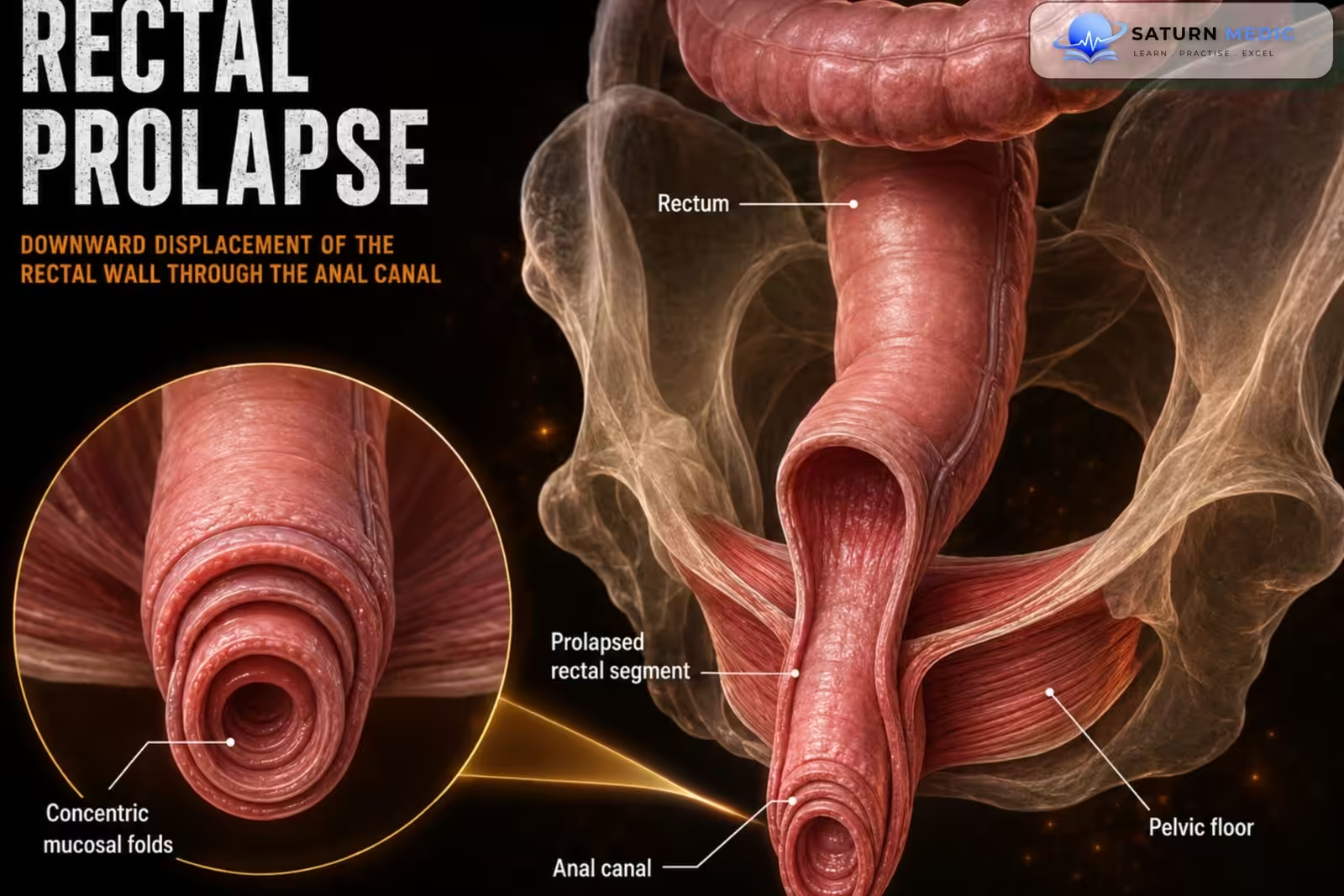
Rectal prolapse is the protrusion of the rectal mucosa (mucosal prolapse) or the entire
rectal wall (full-thickness prolapse) through the anal opening
Also called
ICD-10
Specialty
Onset
Reviewed
-
OverviewOverview
-
Etiology & Risk FactorsEtiology & Risk Factors
-
PathophysiologyPathophysiology
-
Clinical PresentationClinical Presentation
-
History TakingHistory Taking
-
Physical ExaminationPhysical Examination
-
InvestigationsInvestigations
-
DiagnosisDiagnosis
-
ManagementManagement
-
ComplicationsComplications
-
PrognosisPrognosis
-
Key Points / Clinical PearlsKey Points / Clinical Pearls
Overview
– Mucosal prolapse is more common in children.
– Full-thickness rectal prolapse is more common in adults.
– The hallmark presentation is a painless protruding rectal mass during straining.
– Diagnosis is primarily clinical.
– Full-thickness prolapse usually requires surgical treatment.
Etiology & Risk Factors
– Increased intra-abdominal pressure: Excessive straining, Chronic constipation
– Weakness of the pelvic floor muscles: Advanced age, Multiple pregnancies
– Cystic fibrosis (important risk factor in children)
Pathophysiology
Rectal prolapse occurs when:
– The rectal mucosa alone (mucosal prolapse), or
– The entire rectal wall (full-thickness prolapse)
protrudes through the anal canal due to increased intra-abdominal pressure and/or
pelvic floor weakness.
Stretching of the anal sphincter contributes to fecal incontinence
Clinical Presentation
Symptoms
– Painless rectal mass protruding through the anus
– Initially appears only during defecation
– Early stages: Reduces spontaneously after standing
– Later stages: Requires manual (digital) reduction
– Rarely becomes incarcerated: Painful, Cannot be manually reduced
– Fecal incontinence
– Constipation
– Pruritus ani
– Rectal bleeding
History Taking
Assess for: Painless protruding rectal mass, Occurrence during defecation, Need for
manual reduction, Constipation, Fecal incontinence, Rectal bleeding, Pruritus, Duration
and progression, Symptoms of incarceration (pain and inability to reduce)
Physical Examination
– Examine while the patient squats or strains
– Inspect the prolapsed tissue
– Identify: Radial folds (mucosal prolapse), Concentric folds (full-thickness prolapse)
– Assess for: Solitary rectal ulcer, Anal sphincter weakness, Pelvic floor pathology,
Associated pelvic organ prolapse (Uterine prolapse, Vaginal vault prolapse, Cystocele,
Rectocele)
– Perform digital rectal examination
Investigations
Primary investigation
– Clinical examination (usually sufficient)
If diagnosis is uncertain
– Video defecography: Distinguishes full-thickness prolapse from mucosal prolapse;
Barium paste is placed into the rectum and X-rays are taken during defecation
Before surgical treatment
–Proctoscopy and/or colonoscopy: Excludes rectal neoplasm; A rectal tumor may act as a lead point for rectal intussusception
Additional investigations according to presentation
If rectal ulcer present: Biopsy of the ulcer If fecal incontinence present: Anal sphincter manometry
If pelvic floor weakness suspected: Dynamic pelvic floor MRI
Children: Sweat chloride test to exclude cystic fibrosis
Diagnosis
Rectal prolapse is primarily a clinical diagnosis.
Diagnosis is based on: History, Physical examination while straining, Characteristic
appearance of the prolapsed rectum
Related Topics
Management
Mucosal prolapse
– Conservative treatment
– Digital reduction
– Injection sclerotherapy
Full-thickness rectal prolapse
– Usually requires surgery
– Surgical approach: Abdominal approach, or Perineal approach
– Choice of procedure is individualized
Complications
– Fecal incontinence
– Chronic constipation
– Solitary rectal ulcer
– Rectal bleeding
– Incarcerated rectal prolapse (painful and irreducible)
Prognosis
– Mucosal prolapse often responds to conservative treatment.
– Full-thickness rectal prolapse generally requires surgical correction.
– Appropriate treatment usually improves symptoms and quality of life.
Key Points / Clinical Pearls
– Rectal prolapse is primarily a clinical diagnosis.
– Examine the patient while squatting or straining.
– Radial folds = Mucosal prolapse.
– Concentric folds = Full-thickness prolapse.
– Use video defecography when the diagnosis is uncertain.
– Perform proctoscopy and/or colonoscopy before surgery.
– Additional investigations depend on associated findings: Rectal ulcer → Biopsy; Fecal
incontinence → Anal manometry; Pelvic floor weakness → Dynamic pelvic MRI; Children
→ Sweat chloride test
– Mucosal prolapse is managed conservatively or with injection sclerotherapy.
– Full-thickness prolapse usually requires surgery.
- Segal J, McKeown DG, Tavarez MM. National Center for Biotechnology Information (NIH). Rectal Prolapse, StatPearls.
- American Society of Colon and Rectal Surgeons (ASCRS). Treatment of Rectal Prolapse: Clinical Practice Guideline.
- Al Zangana I, Al-Taie RH, Al-Badri S, Ismail M. Rectal Prolapse Surgery: Balancing Effectiveness and Safety in Abdominal and Perineal Approaches. Cureus. 2024;16:e69868. PMID: 39435214.
- MedlinePlus, National Library of Medicine (NIH). Rectal Prolapse: Medical Encyclopedia.
- National Center for Biotechnology Information (NIH). Rectal Prolapse, Surgical Treatment.