

Clinical Subject Page
Pancreatic Pseudocyst
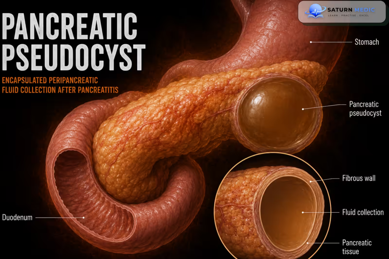
A Pancreatic Pseudocyst is an encapsulated collection of pancreatic fluid that usually develops 4 weeks after an episode of acute pancreatitis and can also occur in chronic pancreatitis. Unlike a true cyst, it does not have an epithelial lining; its wall is made of fibrous (granulation) tissue.
ICD-10
Specialty
Onset
Reviewed
-
OverviewOverview
-
Etiology & Risk FactorsEtiology & Risk Factors
-
PathophysiologyPathophysiology
-
Clinical PresentationClinical Presentation
-
History TakingHistory Taking
-
Physical ExaminationPhysical Examination
-
InvestigationsInvestigations
-
DiagnosisDiagnosis
-
ManagementManagement
-
ComplicationsComplications
-
PrognosisPrognosis
-
Key Points / Clinical PearlsKey Points / Clinical Pearls
Overview
- Forms when pancreatic duct damage causes leakage of pancreatic secretions.
- The leaked fluid triggers inflammation and becomes surrounded by fibrous tissue, forming a pseudocyst.
- Many patients are asymptomatic, while others develop symptoms due to pressure on nearby organs (e.g., abdominal pain, vomiting, jaundice).
Etiology & Risk Factors
Etiology
- Acute pancreatitis (most common cause)
- Chronic pancreatitis
- Pancreatic duct injury or disruption, causing leakage of pancreatic secretions
- The leaked fluid becomes surrounded by granulation (fibrous) tissue, forming a pseudocyst.
Risk Factors
- Previous acute pancreatitis
- Chronic pancreatitis (up to 30% of patients may develop pseudocysts)
- Conditions that increase the risk of pancreatitis (e.g., alcohol-related chronic pancreatitis)
Pancreatic pseudocysts are usually a complication of acute or chronic pancreatitis caused by leakage of pancreatic fluid from damaged pancreatic ducts.
Pathophysiology
Acute/Chronic pancreatitis → Pancreatic duct damage/disruption → Leakage of pancreatic secretions → Inflammatory reaction in surrounding tissue → Encapsulation of fluid by granulation (fibrous) tissue → Formation of a pancreatic pseudocyst
Clinical Presentation
- Often asymptomatic
- Painless abdominal mass
- Pressure effects on nearby organs:
- Gastric outlet obstruction: early satiety, nonbilious vomiting, abdominal pain
- Distal duodenal obstruction: bilious vomiting, steatorrhea
- Bile duct obstruction: jaundice
Most pancreatic pseudocysts are asymptomatic. Symptoms usually occur when the pseudocyst becomes large enough to compress adjacent organs.
History Taking
Ask about:
- Have you had a recent episode of acute pancreatitis? (especially within the last 4 weeks)
- Do you have a history of chronic pancreatitis?
- Do you have persistent or worsening upper abdominal pain?
- Have you noticed a lump or fullness in your upper abdomen?
- Do you feel full quickly (early satiety) or have nausea/vomiting?
- Is the vomiting bilious (green) or nonbilious?
- Have you developed jaundice (yellow eyes/skin)?
- Have you had unexplained weight loss or loss of appetite?
- Do you have fever or chills? (suggests infection)
- Have you had sudden severe abdominal pain, dizziness, or fainting? (suggests rupture or hemorrhage)
Physical Examination
General examination
- Usually normal if asymptomatic
- Jaundice if bile duct obstruction is present
- Fever may indicate an infected pseudocyst
Abdominal examination
- Painless, palpable upper abdominal (epigastric) mass
- Epigastric fullness or distention
- Mild abdominal tenderness if symptomatic
- Signs of gastric outlet obstruction (abdominal distention) in large pseudocysts
Signs of complications
- Jaundice → biliary obstruction
- Fever and abdominal pain → infected pseudocyst
- Sudden severe abdominal pain with signs of shock → hemorrhage or rupture (medical emergency)
Investigations
Imaging (Main investigations)
- CT abdomen with IV contrast – First-line and preferred test
- Shows a well-defined, encapsulated extrapancreatic fluid collection
- Abdominal ultrasound
- Fast and readily available
- Useful for detecting pseudocysts but may miss small lesions
- MRI/MRCP
- Highly sensitive and specific
- Better delineates pancreatic duct anatomy
- ERCP
- More invasive; mainly used when therapeutic intervention (e.g., drainage) is planned
Laboratory tests
- Routine blood tests are used to assess the underlying pancreatitis and complications (no laboratory test specifically confirms a pancreatic pseudocyst).
Contrast-enhanced CT is the investigation of choice for diagnosing and assessing a pancreatic pseudocyst.
Diagnosis
Diagnosis is based on :
Diagnostic Features
- History of acute or chronic pancreatitis (typically ≥ 4 weeks after acute pancreatitis)
- Contrast-enhanced CT abdomen (first-line)
- Shows an extrapancreatic fluid collection
- Well-defined wall/capsule with contrast enhancement
- Supportive imaging (if needed)
- Abdominal ultrasound
- MRI/MRCP
- ERCP (gold standard when therapeutic intervention is planned)
-A well-encapsulated pancreatic fluid collection seen on contrast CT ≥ 4 weeks after pancreatitis is diagnostic of a pancreatic pseudocyst.
Related Topics
Management
1. Conservative management
(for small ≤ 5 cm, asymptomatic, uncomplicated pseudocysts)
- Observation with imaging follow-up
- Pain control (nonopioid analgesics if possible)
- Antiemetics if needed
- Low-fat diet with small, frequent meals
- Alcohol cessation
2. Endoscopic drainage (First-line intervention)
Indications:
- Symptomatic pseudocysts (persistent pain, nausea/vomiting, anorexia, weight loss)
- Large (> 5 cm) and persistent (> 6 weeks) pseudocysts
- Complications:
- Infection
- Hemorrhage
- Gastric outlet, duodenal, or biliary obstruction
- Pancreatic pseudoaneurysm
- Compression of major vessels
- Suspected malignancy
3. Percutaneous drainage
- Used if endoscopic drainage is not feasible or has failed
4. Surgical drainage
- Reserved for failed endoscopic/percutaneous treatment or when surgery is needed for associated complications
- Procedures include:
- Cystogastrostomy
- Cystoduodenostomy
- Cystojejunostomy
Complications
- Infection → fever, abdominal pain, sepsis
- Rupture → pancreatic ascites, pancreaticopleural fistula
- Hemorrhage (erosion into adjacent blood vessels) → sudden severe abdominal pain, hemorrhagic shock
- Gastric outlet obstruction → early satiety, nausea, nonbilious vomiting
- Duodenal obstruction → bilious vomiting, steatorrhea
- Biliary obstruction → obstructive jaundice
- Pancreatic pseudoaneurysm
- Compression of major blood vessels
- Pancreaticopleural fistula
Prognosis
- Good prognosis in most patients with small (≤ 5 cm), asymptomatic pseudocysts, as many resolve spontaneously with conservative management.
- Large (> 5 cm), persistent (> 6 weeks), symptomatic, or complicated pseudocysts are unlikely to resolve without intervention and usually require drainage.
- Endoscopic drainage has a high success rate and is the preferred treatment when intervention is needed.
- Prognosis worsens if complications develop, such as infection, hemorrhage, or rupture, which require urgent management.
Key Points / Clinical Pearls
- Occurs ≥ 4 weeks after pancreatitis.
- No epithelial lining (fibrous wall only).
- Best test: Contrast-enhanced CT abdomen.
- Small (≤ 5 cm), asymptomatic → observe.
- Large (> 5 cm), persistent (> 6 weeks), symptomatic, or complicated → endoscopic drainage.
- Major complications: Infection, hemorrhage, rupture, and obstruction.
- National Center for Biotechnology Information (NIH). Pancreatic Pseudocyst, StatPearls.
- Banks PA, Bollen TL, Dervenis C, et al; Acute Pancreatitis Classification Working Group. Classification of Acute Pancreatitis - 2012: Revision of the Atlanta Classification and Definitions by International Consensus. Gut. 2013;62:102-111.
- Pancreatic Pseudocyst: The Past, the Present, and the Future. World J Gastroenterol. PMC11287700.
- Systematic Review Comparing Endoscopic, Percutaneous and Surgical Pancreatic Pseudocyst Drainage. World J Gastroenterol. PMC4804189.
- MedlinePlus, National Library of Medicine (NIH). Pancreatic Pseudocyst: Medical Encyclopedia.