

Clinical Subject Page
Liver Cirrhosis
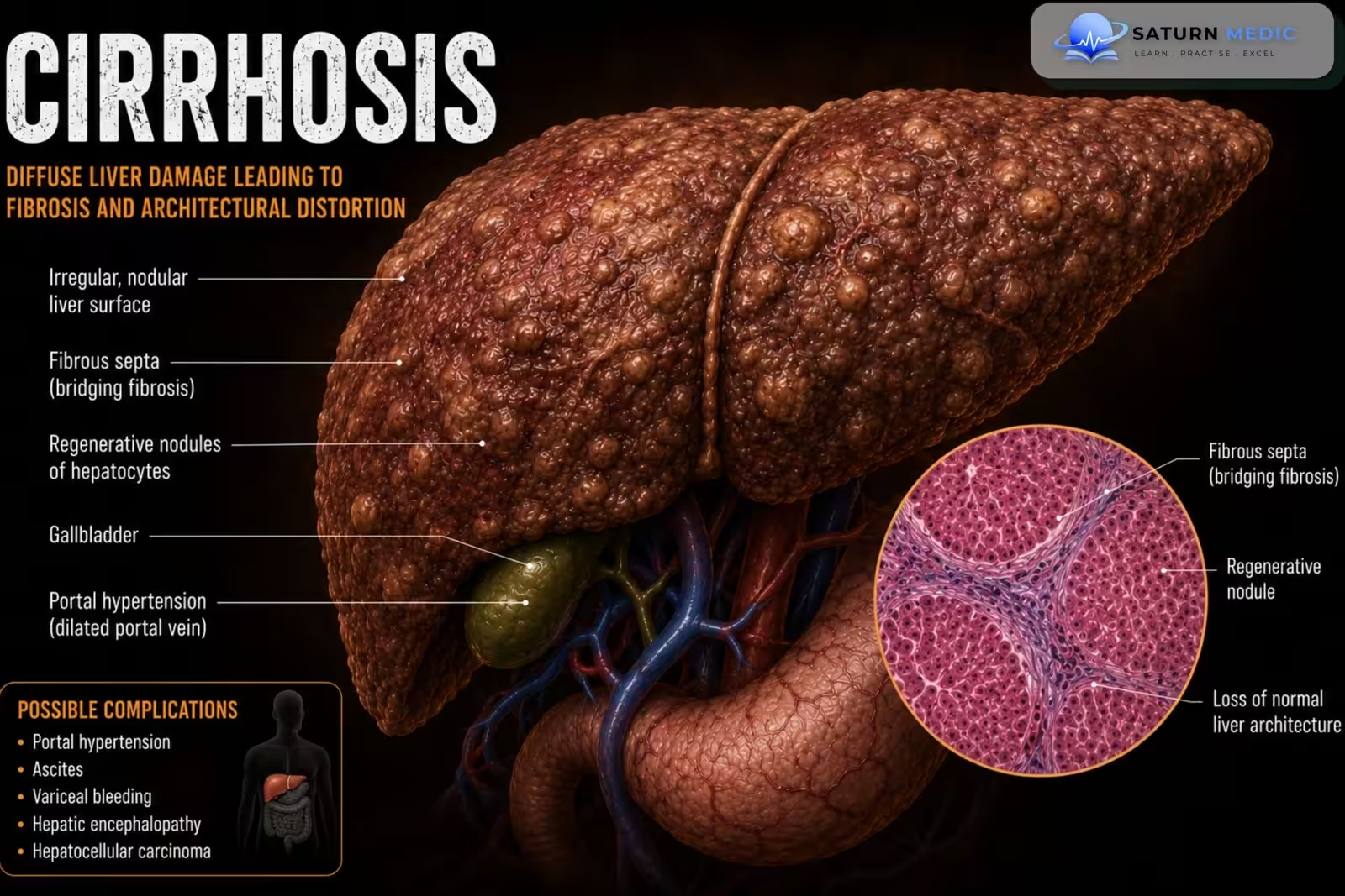
Liver cirrhosis is the end stage of chronic liver disease characterized by diffuse hepatic fibrosis and
regenerative nodules, resulting in impaired liver function and portal hypertension.
ICD-10
Specialty
Onset
Reviewed
-
OverviewOverview
-
Etiology & Risk FactorsEtiology & Risk Factors
-
PathophysiologyPathophysiology
-
Clinical PresentationClinical Presentation
-
History TakingHistory Taking
-
Physical ExaminationPhysical Examination
-
InvestigationsInvestigations
-
DiagnosisDiagnosis
-
ManagementManagement
-
ComplicationsComplications
-
PrognosisPrognosis
-
Key Points / Clinical PearlsKey Points / Clinical Pearls
Overview
- • Progressive fibrosis replaces normal liver architecture.
• Initially patients may have compensated disease with few or no symptoms.
• Decompensated cirrhosis presents with complications such as ascites, variceal bleeding, hepatic
encephalopathy, and jaundice.
• Major causes include chronic hepatitis B, chronic hepatitis C, alcohol-associated liver disease,
MASLD (NAFLD), autoimmune hepatitis, and hereditary liver diseases.
Etiology & Risk Factors
• Chronic hepatitis B
• Chronic hepatitis C
• Alcohol-associated liver disease
• Metabolic dysfunction-associated steatotic liver disease (MASLD)
• Autoimmune hepatitis
• Hemochromatosis
• Wilson disease
• Alpha-1 antitrypsin deficiency
• Primary biliary cholangitis
• Primary sclerosing cholangitis
Pathophysiology
• Chronic liver injury causes persistent inflammation.
• Progressive fibrosis develops.
• Regenerative nodules distort normal liver architecture.
• Portal hypertension develops.
• Hepatic synthetic and metabolic functions progressively decline.
Clinical Presentation
-Compensated cirrhosis:
- Often asymptomatic or discovered incidentally.
-Decompensated cirrhosis:
• Fatigue
• Jaundice
• Ascites
• Peripheral edema
• Gastrointestinal bleeding
• Hepatic encephalopathy
• Pruritus
• Weight loss
History Taking
-Ask about:
• Alcohol consumption
• Viral hepatitis risk factors
• Previous liver disease
• Family history
• Drug history
• Features of chronic liver disease
• Hematemesis or melena
• Abdominal distension
• Confusion or altered mental status
• Weight loss
• Pruritus
Physical Examination
• Jaundice
• Hepatomegaly or small shrunken liver
• Splenomegaly
• Ascites
• Peripheral edema
• Spider angiomas
• Palmar erythema
• Caput medusae
• Muscle wasting
• Asterixis
• Signs of hepatic encephalopathy
Investigations
1. Routine laboratory studies
Liver chemistry
• ALT • AST • Bilirubin • ALP • GGT
Liver synthetic function
• PT/INR • Albumin • Total protein
Complete blood count
• Thrombocytopenia • Anemia • Leukopenia
Electrolytes/CMP
• Hyponatremia
2. Investigate the underlying cause
Viral hepatitis: HBsAg, IgM anti-HBc, Anti-HCV, IgM HAV, IgM HEV
-MASLD: HbA1c, Fasting lipid profile
-Hemochromatosis: Serum iron, Ferritin, Transferrin saturation
-Autoimmune hepatitis: ANA, ASMA, Anti-LKM-1, Anti-soluble liver antigen antibody, Total IgG
-Wilson disease: Ceruloplasmin, Serum copper, Urinary copper
Alpha-1 antitrypsin deficiency: Alpha-1 antitrypsin level and phenotype
-Primary biliary cholangitis: AMA (AMA-M2), ALP, Bilirubin
Primary sclerosing cholangitis: ALP, GGT, Bilirubin, pANCA, IgG
3. Imaging
Abdominal ultrasound with Doppler (Best initial imaging): Nodular liver surface, Liver size,
Fibrosis, Portal hypertension, Splenomegaly, Ascites, Portal vein thrombosis, Hepatocellular
carcinoma surveillance.
• CT abdomen if ultrasound is inadequate.
4. Noninvasive fibrosis assessment
• Liver elastography (FibroScan®) • APRI score • FIB-4 score
5. Liver biopsy
Indications: Diagnostic uncertainty, Determine etiology, Grade and stage fibrosis, Evaluate focal
liver lesions.
Diagnosis
-Diagnosis is based on:
• Clinical features
• Laboratory abnormalities
• Imaging findings
• Fibrosis assessment
• Liver biopsy when required
Related Topics
Management
-General management
• Treat the underlying cause.
• Avoid alcohol.
• Avoid hepatotoxic medications.
• Ensure appropriate vaccinations.
• Optimize nutrition (adequate calories and protein).
• Regular follow-up and monitoring.
-Management of complications
Ascites: Sodium restriction (<2 g/day), Spironolactone + furosemide, Large-volume paracentesis
with albumin for tense ascites.
-Hepatic encephalopathy: Lactulose, Add rifaximin for recurrent or persistent encephalopathy.
-Variceal bleeding: Urgent upper endoscopy, Octreotide infusion, Ceftriaxone prophylaxis,
Carvedilol for prevention of variceal bleeding.
-Suspected spontaneous bacterial peritonitis (SBP): Cefotaxime, Ascitic fluid analysis.
Monitoring
• CBC
• Liver function tests
• INR
• Renal function and electrolytes
• Abdominal ultrasound for
hepatocellular carcinoma surveillance
Liver transplantation
Refer for transplant evaluation in: Decompensated cirrhosis; MELD score ≥15
Complications
• Portal hypertension
• Ascites
• Spontaneous bacterial peritonitis
• Esophageal varices
• Variceal hemorrhage
• Hepatic encephalopathy
• Hepatorenal syndrome
• Portal vein thrombosis
• Hepatocellular carcinoma
• Liver failure
Prognosis
• Depends on the stage of disease and underlying cause.
• Compensated cirrhosis has a better prognosis.
• Development of decompensation significantly worsens survival.
• Liver transplantation improves outcomes in selected patients.
Key Points / Clinical Pearls
• Cirrhosis is the end stage of chronic liver disease.
• Diagnosis combines clinical features, laboratory tests, imaging, and fibrosis assessment.
• Abdominal ultrasound with Doppler is the best initial imaging study.
• FibroScan®, APRI, and FIB-4 help assess liver fibrosis.
• Management focuses on treating the underlying cause and preventing complications.
• Ascites → sodium restriction + spironolactone/furosemide ± paracentesis.
• Hepatic encephalopathy → lactulose ± rifaximin.
• Variceal bleeding → octreotide + ceftriaxone + urgent endoscopy.
• Regular HCC surveillance is essential.
• Liver transplantation is indicated for decompensated cirrhosis or MELD ≥15.
- National Center for Biotechnology Information (NIH). Chronic Liver Disease, StatPearls.
- Tsoris A, Marlar CA. National Center for Biotechnology Information (NIH). Use of the Child Pugh Score in Liver Disease, StatPearls.
- Kim WR, Mannalithara A, Heimbach JK, et al. MELD 3.0: The Model for End-Stage Liver Disease Updated for the Modern Era. Gastroenterology. 2021;161:1887-1895.
- MedlinePlus, National Library of Medicine (NIH). Cirrhosis: Health Topic.
- National Center for Biotechnology Information (NIH). Portal Hypertension, StatPearls.