

Clinical Subject Page
Acute Cholecystitis
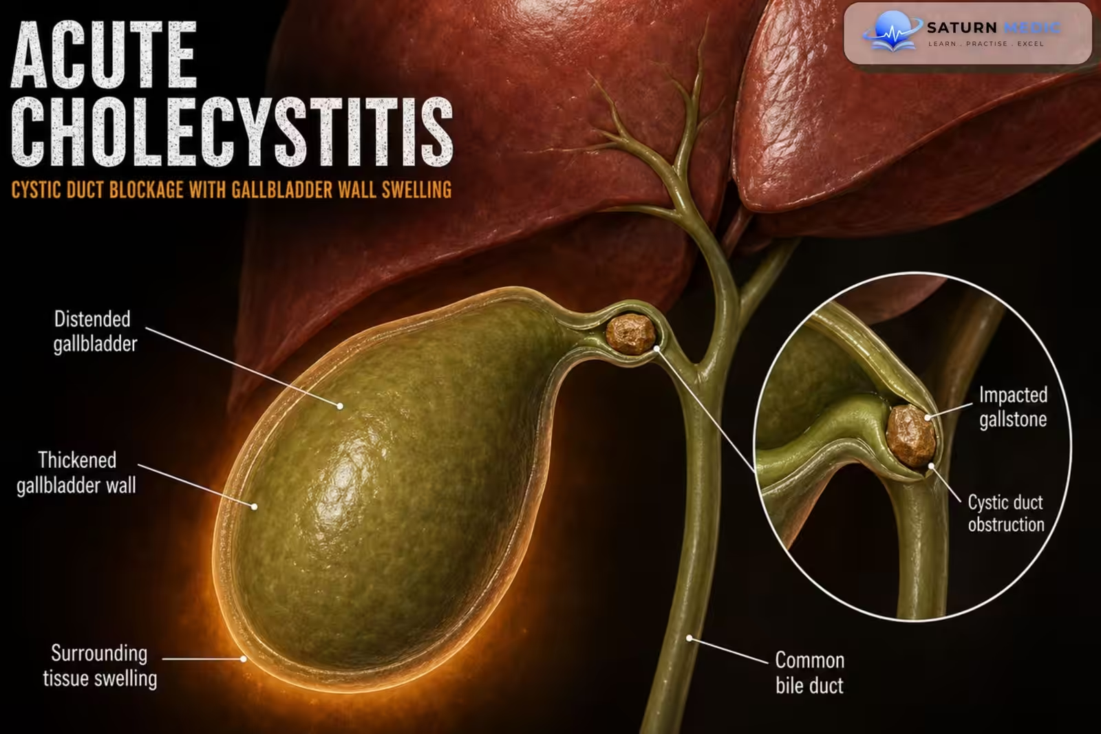
Acute cholecystitis is acute inflammation of the gallbladder, most commonly caused by obstruction
of the cystic duct by a gallstone (acute calculous cholecystitis). Acalculous cholecystitis occurs
mainly in critically ill patients.
Also called
ICD-10
Specialty
Onset
Reviewed
-
OverviewOverview
-
Etiology & Risk FactorsEtiology & Risk Factors
-
PathophysiologyPathophysiology
-
Clinical PresentationClinical Presentation
-
History TakingHistory Taking
-
Physical ExaminationPhysical Examination
-
InvestigationsInvestigations
-
DiagnosisDiagnosis
-
ManagementManagement
-
ComplicationsComplications
-
PrognosisPrognosis
-
Key Points / Clinical PearlsKey Points / Clinical Pearls
Overview
• Usually results from gallstone obstruction of the cystic duct.
• Characterized by persistent right upper quadrant (RUQ) pain, Murphy sign, and systemic
inflammation.
• RUQ ultrasound is the preferred initial imaging modality.
• Main treatment includes supportive care, empiric antibiotics, and early laparoscopic
cholecystectomy.
Etiology & Risk Factors
-Causes:
• Acute calculous cholecystitis (most common): Cystic duct obstruction by a gallstone.
• Acute acalculous cholecystitis: Typically occurs in critically ill patients.
Risk Factors
• Gallstones
• Critical illness (for acalculous cholecystitis)
Pathophysiology
• Obstruction of the cystic duct leads to gallbladder distension.
• Inflammation and edema develop within the gallbladder wall.
• Secondary bacterial infection may occur.
• Untreated disease may progress to gangrene, perforation, or abscess formation.
Clinical Presentation
• Persistent right upper quadrant pain
• Fever
• Nausea
• Vomiting
• Positive Murphy sign
History Taking
-Ask about:
• Onset and duration of RUQ pain
• Pain severity
• Fever
• Nausea and vomiting
• Previous gallstone attacks
• Jaundice
• History of gallstones
• Previous biliary surgery
Physical Examination
• General assessment (ABCDE)
• Vital signs
• Right upper quadrant tenderness
• Murphy sign
• Assess for jaundice
• Evaluate for signs of sepsis or shock
Investigations
Initial laboratory studies
• Complete blood count (CBC)
• Basic metabolic panel (BMP)
• C-reactive protein (CRP)
• Coagulation studies
-Liver chemistries
• Lipase
• Amylase
-Blood cultures (2 sets) before antibiotics if infection is suspected
Imaging
Preferred initial imaging
• Right upper quadrant (RUQ) ultrasound
• Consider biliary point-of-care ultrasound (POCUS) if available.
Typical ultrasound findings:
• Gallbladder distension
• Gallbladder wall edema/thickening
• Pericholecystic fluid
Additional imaging (if ultrasound is inconclusive)
• CT abdomen
• MRI abdomen
• MRCP
-HIDA scan:
Evaluate for
• Choledocholithiasis
• Severity of acute cholecystitis
Diagnosis
-Diagnosis is based on:
1) Local signs of inflammation
• Murphy sign
• RUQ pain/tenderness
2) Systemic signs of inflammation
• Fever
• Leukocytosis
• Elevated CRP
3) Imaging
• Characteristic evidence of gallbladder inflammation on imaging
Related Topics
Management
-Initial management
• Admit to hospital (all patients require inpatient management)
• Establish IV access
• Hemodynamic support
• Nil by mouth (NPO)
• IV fluids
• Analgesia
• Antiemetics
• Consider NG tube for intractable vomiting
-Antibiotics
• Start empiric antibiotic therapy for acute biliary infection promptly.
-Surgical management
Definitive treatment
• Early laparoscopic cholecystectomy
• Preferably performed within 72 hours of admission
-High-risk surgical patients
• Gallbladder drainage (e.g., percutaneous cholecystostomy or endoscopic gallbladder
stenting)
• Elective interval cholecystectomy after recovery
Consultations
• General surgery consultation is recommended for all patients
Complications
• Gangrenous cholecystitis
• Emphysematous cholecystitis
• Gallbladder perforation
• Biliary-enteric fistula
• Gallstone ileus
• Pyogenic liver abscess
• Chronic cholecystitis
• Increased risk of gallbladder carcinoma
Prognosis
• Excellent with early diagnosis and treatment.
• Delayed management increases the risk of complications.
• Early laparoscopic cholecystectomy reduces recurrence and improves outcomes.
Key Points / Clinical Pearls
• Acute cholecystitis is usually caused by cystic duct obstruction from a gallstone.
• Classic features: RUQ pain, Murphy sign, and fever.
• RUQ ultrasound is the investigation of choice.
• Initial labs: CBC, BMP, CRP, liver chemistries, amylase, lipase, coagulation studies, and
blood cultures.
• Diagnosis requires local signs of inflammation + systemic inflammation + imaging findings.
• Management includes NPO, IV fluids, analgesia, empiric antibiotics, and early laparoscopic
cholecystectomy.
• High-risk patients may require percutaneous cholecystostomy before definitive surgery
- National Center for Biotechnology Information (NIH). Acute Cholecystitis, StatPearls.
- Yokoe M, Hata J, Takada T, et al. Tokyo Guidelines 2018: Diagnostic Criteria and Severity Grading of Acute Cholecystitis (with videos). J Hepatobiliary Pancreat Sci. 2018;25:41-54. DOI: 10.1002/jhbp.515.
- Mori Y, Itoi T, Baron TH, et al. Tokyo Guidelines 2018: Management Strategies for Gallbladder Drainage in Patients With Acute Cholecystitis. J Hepatobiliary Pancreat Sci. 2018;25:87-95. Tokyo Guidelines Flowchart.
- MedlinePlus, National Library of Medicine (NIH). Acute Cholecystitis: Medical Encyclopedia.
- National Center for Biotechnology Information (NIH). Gallstones (Cholelithiasis), StatPearls.