

Clinical Subject Page
Choledocholithiasis
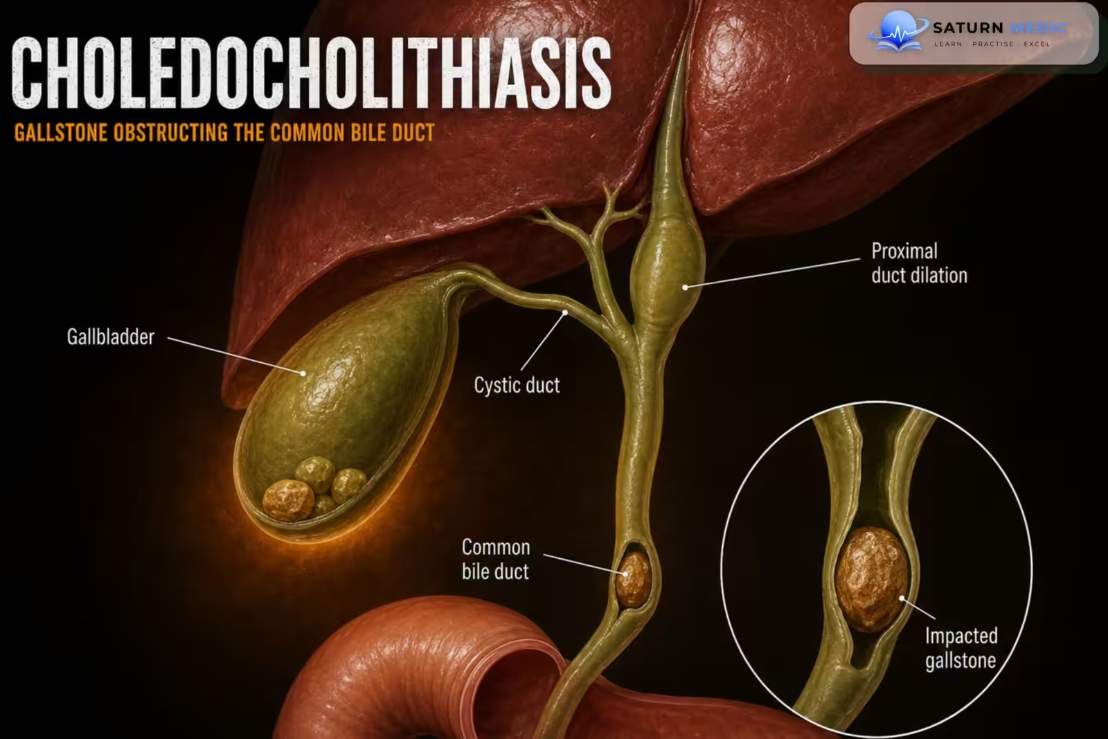
Choledocholithiasis is the presence of gallstones within the common bile duct (CBD). It may
cause biliary obstruction and can lead to serious complications such as acute cholangitis and
gallstone pancreatitis
Also called
ICD-10
Specialty
Onset
Reviewed
-
OverviewOverview
-
Etiology & Risk FactorsEtiology & Risk Factors
-
PathophysiologyPathophysiology
-
Clinical PresentationClinical Presentation
-
History TakingHistory Taking
-
Physical ExaminationPhysical Examination
-
InvestigationsInvestigations
-
DiagnosisDiagnosis
-
ManagementManagement
-
ComplicationsComplications
-
PrognosisPrognosis
-
Key Points / Clinical PearlsKey Points / Clinical Pearls
Overview
• Gallstones migrate from the gallbladder into the CBD.
• Patients commonly present with right upper quadrant pain and features of extrahepatic
cholestasis.
• Liver function tests (LFTs) and RUQ ultrasound are the preferred first-line investigations.
• Confirmatory imaging depends on the likelihood of choledocholithiasis.
• Treatment involves stone removal (usually ERCP) and prevention of recurrence with
laparoscopic cholecystectomy.
Etiology & Risk Factors
-Cause
• Gallstones within the common bile duct (CBD), usually migrating from the gallbladder.
-Risk factors
• Symptomatic cholelithiasis
• Previous gallstones
Pathophysiology
• A gallstone obstructs the common bile duct.
• Biliary obstruction causes cholestasis.
• Persistent obstruction may result in:
• Acute cholangitis
• Gallstone pancreatitis
• Obstructive jaundice
Clinical Presentation
-Typical features include:
• Right upper quadrant pain
• Jaundice
• Signs of extrahepatic cholestasis
Assess for complications:
• Acute cholangitis
• Acute pancreatitis
• Acute cholecystitis
History Taking
-Ask about:
• Right upper quadrant pain
• Jaundice
• Previous gallstones
• Previous biliary surgery
• Fever or chills
• Symptoms suggestive of pancreatitis
• Symptoms suggestive of cholangitis
Physical Examination
• ABCDE assessment
• Vital signs
• Right upper quadrant tenderness
• Assess for jaundice
• Look for signs of cholangitis
• Evaluate for pancreatitis and cholecystitis
Investigations
Initial laboratory studies
• Complete blood count (CBC)
• Liver chemistries (LFTs)
• Amylase
• Lipase
Initial imaging
Preferred first-line investigations
• Right upper quadrant (RUQ) ultrasound
• Biliary point-of-care ultrasound (POCUS) (if available)
-CBD stones are rarely visualized on ultrasound.
Normal LFTs and ultrasound may help rule out
disease but cannot reliably confirm the diagnosis.
-Confirmatory imaging (based on risk)
•High likelihood
• ERCP
Intermediate likelihood
• MRCP
• Endoscopic ultrasound (EUS)
•Low likelihood
• Confirmatory imaging is not routinely required
Risk stratification
-Very strong predictors
• CBD stone seen on ultrasound
• Clinical signs of cholangitis
• Total bilirubin >4 mg/dL
Strong predictors
• Dilated CBD (>6 mm with gallbladder in situ)
• Total bilirubin 1.8–4 mg/dL
-Moderate predictors
• Abnormal liver tests (other than bilirubin)
• Age >55 years
• Clinical features of biliary pancreatitis
Diagnosis
Diagnosis is based on:
• Clinical presentation
• Liver function tests
• RUQ ultrasound
• Risk stratification
• ERCP, MRCP, or EUS when indicated
Related Topics
Management
-Initial management
• Nil by mouth (NPO)
• Analgesia
• Antiemetics
• Supportive care for acute biliary disease
-Definitive management
High likelihood of choledocholithiasis
• Consult gastroenterology
• ERCP for diagnosis and stone removal
Intermediate likelihood
• MRCP or EUS followed by appropriate intervention
-Treat associated complications Manage if present:
• Acute cholangitis
• Acute pancreatitis
• Acute cholecystitis
Prevention of recurrence
• Laparoscopic cholecystectomy after stone clearance when appropriate
Complications
• Acute cholangitis
• Gallstone pancreatitis
• Obstructive jaundice
• Persistent biliary obstruction
Prognosis
• Excellent with timely diagnosis and stone removal.
• Delayed treatment increases the risk of cholangitis, pancreatitis, and other complications.
• Cholecystectomy reduces the risk of recurrent biliary events.
Key Points / Clinical Pearls
• Choledocholithiasis = gallstones in the common bile duct.
• First-line investigations: LFTs + RUQ ultrasound.
• Initial labs: CBC, liver chemistries, amylase, and lipase.
• ERCP is indicated for patients with a high likelihood of choledocholithiasis and allows stone
removal.
• MRCP or EUS are preferred for patients with an intermediate likelihood.
• Always assess for acute cholangitis, acute pancreatitis, and acute cholecystitis.
• Definitive management includes stone extraction and laparoscopic cholecystectomy to
prevent recurrence.
- McNicoll CF, Pastorino A, Farooq U, Froehlich MJ, St Hill CR. National Center for Biotechnology Information (NIH). Choledocholithiasis, StatPearls.
- ASGE Standards of Practice Committee, Buxbaum JL, Abbas Fehmi SM, Sultan S, et al. ASGE Guideline on the Role of Endoscopy in the Evaluation and Management of Choledocholithiasis. Gastrointest Endosc. 2019;89:1075-1105. ASGE Guideline.
- Manes G, Paspatis G, Aabakken L, et al. Endoscopic Management of Common Bile Duct Stones: European Society of Gastrointestinal Endoscopy (ESGE) Guideline. Endoscopy. 2019;51:472-491. ESGE Guideline.
- Baiu I, Hawn MT. Choledocholithiasis. JAMA. 2018;320:1506. DOI: 10.1001/jama.2018.11812.
- MedlinePlus, National Library of Medicine (NIH). Bile Duct Obstruction: Medical Encyclopedia.