

Clinical Subject Page
Gallbladder Carcinoma
Gallbladder carcinoma is the most common malignancy of the biliary tract, originating from the
mucosal lining of the gallbladder. It is often diagnosed incidentally after cholecystectomy and is
usually advanced at presentation.
Also called
ICD-10
Specialty
Onset
Reviewed
-
OverviewOverview
-
Etiology & Risk FactorsEtiology & Risk Factors
-
PathophysiologyPathophysiology
-
Clinical PresentationClinical Presentation
-
History TakingHistory Taking
-
Physical ExaminationPhysical Examination
-
InvestigationsInvestigations
-
DiagnosisDiagnosis
-
ManagementManagement
-
ComplicationsComplications
-
PrognosisPrognosis
-
Key Points / Clinical PearlsKey Points / Clinical Pearls
Overview
• Rare but highly aggressive malignancy.
• Most cases are adenocarcinomas.
• Chronic gallbladder inflammation is the major predisposing factor.
• Early disease is often asymptomatic.
• Management depends on tumor stage and resectability.
Etiology & Risk Factors
• Cholelithiasis (most common risk factor)
• Chronic cholecystitis
• Porcelain gallbladder
• Choledocholithiasis
• Chronic cholangitis (e.g., salmonellosis)
• Gallbladder polyps
• Liver fluke infection (e.g., Clonorchis sinensis)
Pathophysiology
• Chronic inflammation of the gallbladder promotes malignant transformation of the mucosal
epithelium.
• Most tumors are adenocarcinomas.
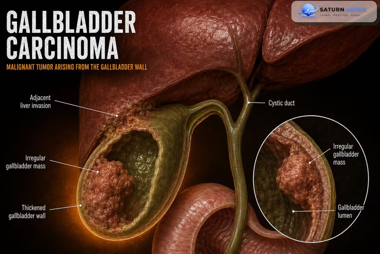
• Tumors may invade the liver, lymph nodes, and adjacent structures.
Clinical Presentation
Early disease
• Usually asymptomatic
Symptomatic disease
• Right upper quadrant or epigastric pain
• Symptoms resembling biliary colic or chronic cholecystitis
• Weight loss
• Nausea
• Weakness
• Fatigue
• Abdominal mass
Advanced disease
• Obstructive jaundice (may occur, sometimes with Courvoisier sign)
History Taking
-Ask about:
• Right upper quadrant pain
• Weight loss
• Loss of appetite
• Nausea
• Fatigue
• History of gallstones
• Chronic cholecystitis
• Gallbladder polyps
• Previous biliary disease
Physical Examination
• General examination
• Right upper quadrant tenderness
• Abdominal mass
• Jaundice (advanced disease)
• Signs of biliary obstruction
• Assessment for metastatic disease
Investigations
Laboratory studies
• Liver chemistries (often normal)
• Tumor markers: CA 19-9, CEA
Tumor markers are used for baseline monitoring, not for diagnosis.
Imaging
• Transabdominal ultrasound: Gallbladder mass, Gallbladder wall thickening
• MRI/MRCP (preferred for evaluating local disease and vascular involvement)
• Multidetector CT (MDCT) abdomen and pelvis
• CT chest (± whole-body PET-CT) for staging
• Endoscopic ultrasound (EUS): Better visualization; may allow tissue sampling when required.
Pathology
• Preoperative biopsy is usually unnecessary.
• Diagnosis is commonly established from the resected surgical specimen.
• Adenocarcinoma is the most common histological type.
-Molecular testing: Consider in advanced disease or high risk of recurrence to identify therapeutic
targets (e.g., HER2 expression, IDH1 mutation).
Diagnosis
-Diagnosis is based on:
• Clinical suspicion
• Imaging studies
• Histopathology after surgical resection
• Staging with cross-sectional imaging
Related Topics
Management
-Surgical management (Curative treatment)
•T1a disease: Simple cholecystectomy may be sufficient.
•≥T1b disease: Extended surgical resection – Liver tissue resection; Portal lymph node dissection.
•Severe obstructive jaundice: Preoperative percutaneous transhepatic biliary drainage may be performed.
-Adjuvant therapy
After resection of T2 or more advanced disease: Gemcitabine + Cisplatin.
-Unresectable or advanced disease
• First-line: Gemcitabine + Cisplatin
• Second-line: FOLFOX; Targeted therapy when appropriate based on molecular testing
• Radiotherapy: May be used for palliative treatment.
Complications
• Local liver invasion
• Lymph node metastasis
• Biliary obstruction
• Metastatic disease
• Death
Prognosis
• Prognosis is generally poor.
• 5-year survival is <5% overall.
• Early-stage disease treated surgically offers the best chance of cure.
Key Points / Clinical Pearls
• Gallbladder carcinoma is the most common biliary tract malignancy.
• Cholelithiasis is the strongest risk factor.
• Early disease is often asymptomatic.
• Transabdominal ultrasound is the initial imaging study.
• MRI/MRCP is preferred for local staging.
• CT chest ± PET-CT is used for staging.
• CA 19-9 and CEA are monitoring markers, not diagnostic tests.
• Preoperative biopsy is usually unnecessary.
• Simple cholecystectomy is adequate for T1a disease.
• Extended resection is required for ≥T1b disease.
• Advanced disease is treated with gemcitabine + cisplatin, with FOLFOX or targeted therapy as
later options
- National Center for Biotechnology Information (NIH). Gallbladder Carcinoma, StatPearls.
- National Comprehensive Cancer Network (NCCN). NCCN Clinical Practice Guidelines in Oncology: Biliary Tract Cancers, Version 1.2026.
- Machado NO. Porcelain Gallbladder: Decoding the Malignant Truth. Sultan Qaboos Univ Med J. 2016;16:e416-e421. PMC5135451.
- National Center for Biotechnology Information (NIH). Porcelain Gallbladder, StatPearls.
- MedlinePlus, National Library of Medicine (NIH). Gallbladder Cancer: Health Topic.