

Clinical Subject Page
Pheochromocytoma
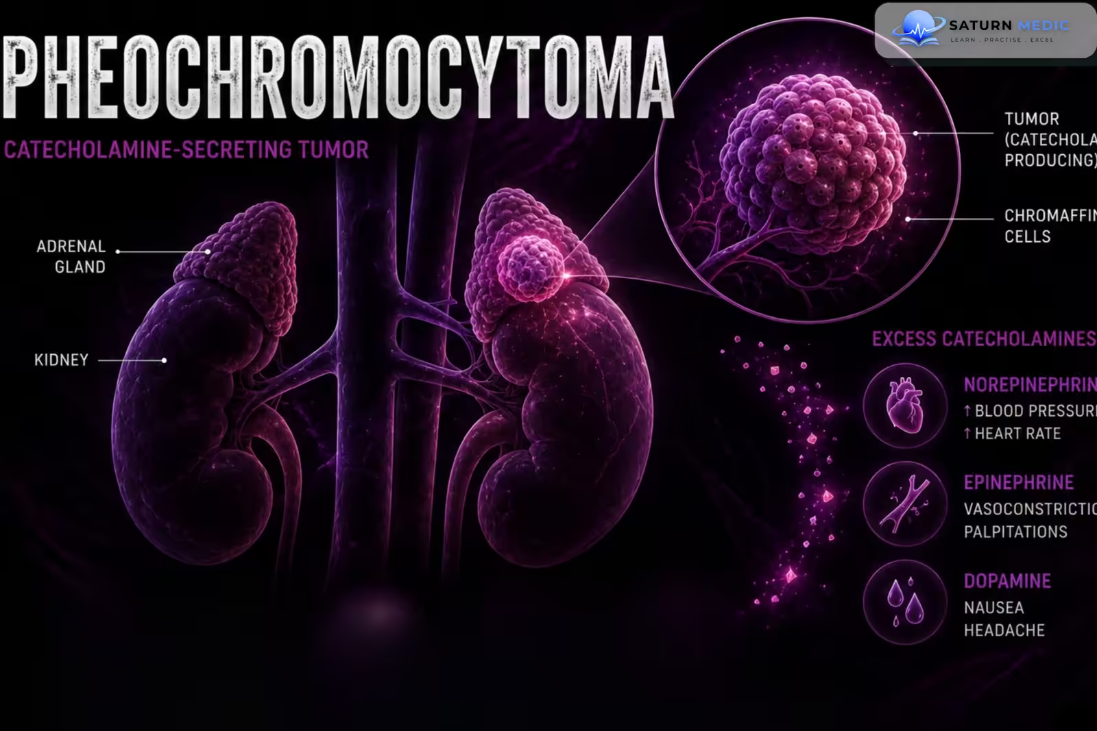
Pheochromocytoma is a catecholamine-producing tumor arising from chromaffin cells, usually in the adrenal medulla. It causes excessive secretion of epinephrine, norepinephrine, or both, often resulting in episodic or persistent hypertension.
Also called
Chromaffin Cell Tumor
ICD-10
D35.00
Specialty
Endocrine
Onset
episodic/paroxysmal
Reviewed
August 2026
On This Page
-
OverviewOverview
-
Etiology & Risk FactorsEtiology & Risk Factors
-
PathophysiologyPathophysiology
-
Clinical PresentationClinical Presentation
-
History TakingHistory Taking
-
Physical ExaminationPhysical Examination
-
InvestigationsInvestigations
-
DiagnosisDiagnosis
-
ManagementManagement
-
ComplicationsComplications
-
PrognosisPrognosis
-
Key Points / Clinical PearlsKey Points / Clinical Pearls
Overview
–Pheochromocytoma is a rare neuroendocrine tumor that produces excessive catecholamines.
-Classic clinical features include:
-
-
- Episodic headache
- Sweating
- Palpitations
- Hypertension
- The classic triad is highly suggestive but is not present in every patient.
-
Etiology & Risk Factors
-Etiology:
-Pheochromocytoma arises from chromaffin cells of the adrenal medulla.
Catecholamine secretion may include:
- Norepinephrine
- Epinephrine
- Dopamine
Most tumors are located in the adrenal glands, while related extra-adrenal tumors are called paragangliomas.
-A significant proportion of pheochromocytomas are associated with hereditary syndromes or pathogenic germline variants.
-Important associations include:
- Multiple Endocrine Neoplasia type 2 (MEN2)
- Von Hippel-Lindau disease (VHL)
- Neurofibromatosis type 1 (NF1)
-Risk Factors:
- Family history of pheochromocytoma or paraganglioma
- MEN2
- Von Hippel-Lindau disease
- Neurofibromatosis type 1
- Hereditary paraganglioma syndromes
Pathophysiology
Chromaffin cell tumor → excessive catecholamine production → α-adrenergic stimulation → vasoconstriction → hypertension
Catecholamine excess → β-adrenergic stimulation → tachycardia + increased cardiac contractility → palpitations
Catecholamine surges → episodic autonomic activation → headache + sweating + palpitations + hypertension
Chronic catecholamine excess → cardiovascular stress → arrhythmias + cardiomyopathy + myocardial injury
Clinical Presentation
-Symptoms:
- Episodic headache
- Excessive sweating
- Palpitations
- Anxiety
- Tremor
- Chest discomfort
- Shortness of breath
- Abdominal pain
- Nausea
- Weight loss
-Signs:
- Hypertension
- Tachycardia
- Orthostatic hypotension
- Pallor
- Tremor
- Excessive sweating
- Weight loss
- Hyperglycemia
History Taking
-Ask about:
- Headaches
- Episodic sweating
- Palpitations
- Blood pressure episodes
- Chest pain
- Shortness of breath
- Tremor
- Anxiety or panic-like episodes
- Weight loss
- Abdominal pain
- Constipation
- Heat intolerance
- Orthostatic dizziness
- Previous hypertensive crises
Physical Examination
–General Examination
- Blood pressure
- Heart rate
- Orthostatic blood pressure
- Body weight
- Hydration status
-Look for:
- Hypertension
- Tachycardia
- Pallor
- Sweating
- Tremor
- Weight loss
-System-Specific Examination:
–Cardiovascular
-Assess for:
- Hypertension
- Tachycardia
- Arrhythmias
- Cardiomyopathy
- Heart failure
–Neurological
-Assess for:
- Headache
- Neurological deficits
- Evidence of previous cerebrovascular complications
–Genetic/Syndromic Examination
-Look for features suggesting:
- Neurofibromatosis type 1
- MEN2
- Von Hippel-Lindau disease
Investigations
-Biochemistry / Specific Tests:
-Plasma Free Metanephrines
A highly sensitive screening test.
-Measure:
- Metanephrine
- Normetanephrine
-24 Hour Urinary Fractionated Metanephrines
An alternative biochemical test.
May also measure:
- Urinary catecholamines
-Additional tests may include:
- Glucose
- Electrolytes
- Renal function
- Liver function
–Imaging
After biochemical evidence of catecholamine excess, localization is usually performed with:
CT or MRI
- Adrenal CT
- Adrenal MRI
- Abdominal/pelvic imaging when extra-adrenal disease is suspected
Diagnosis
-Diagnosis is based on :
Diagnosis requires evidence of excess catecholamine production together with appropriate clinical and/or radiological findings.
-Initial biochemical evaluation
- Plasma free metanephrines
- or
- 24-hour urinary fractionated metanephrines
Markedly elevated results strongly support the diagnosis.
Management
1. First-Line / Emergency Management
-Suspected Pheochromocytoma Crisis
This is a medical emergency.
Management may include:
- Immediate cardiovascular stabilization
- Alpha-adrenergic blockade
- Intravenous fluids when appropriate
- Treatment of severe hypertension
- Management of arrhythmias
- Intensive monitoring
If a patient is not already adequately alpha-blocked, pure beta-blockade should not be started first, because unopposed alpha-adrenergic stimulation can worsen hypertension.
2. Definitive Treatment
Surgical removal of the tumor is the definitive treatment for localized resectable pheochromocytoma.
3. Medical Treatment
Alpha-Adrenergic Blockade
Common options include:
- Phenoxybenzamine
- Selective α1-blockers such as doxazosin
Used preoperatively to:
- Control blood pressure
- Reduce catecholamine-related complications
4. Surgical / Procedural Treatment
- Adrenalectomy
- Usually performed laparoscopically for appropriately selected localized tumors.
- Open surgery may be considered for:
-Before surgery:
- Alpha blockade → volume optimization → beta blockade if needed → tumor resectio
Complications
- Hypertensive crisis
- Cardiac arrhythmias
- Catecholamine-induced cardiomyopathy
- Acute heart failure
- Myocardial ischemia/injury
- Stroke
- Pulmonary edema
- Acute kidney injury
- Hyperglycemia
Prognosis
-The prognosis is generally good for localized tumors that are completely resected.
-Prognosis depends on:
- Tumor location
- Tumor size
- Genetic background
- Completeness of resection
- Presence of metastatic disease
- Recurrence
Key Points / Clinical Pearls
- Pheochromocytoma is a catecholamine-producing tumor of chromaffin cells, usually in the adrenal medulla.
- The classic symptoms are headache, sweating, and palpitations.
- Hypertension may be episodic or persistent.
- Patients may have normal blood pressure between episodes.
- Important hereditary associations include MEN2, von Hippel-Lindau disease, NF1, and hereditary paraganglioma-pheochromocytoma syndromes.
- Plasma free metanephrines or urinary fractionated metanephrines are key biochemical tests.
- Biochemical testing generally comes before localization imaging.
- CT or MRI is used to localize the tumor after biochemical evidence is established.
- Genetic evaluation should be considered in many patients with pheochromocytoma.
- Lenders JWM, Duh QY, Eisenhofer G, et al. Pheochromocytoma and Paraganglioma: An Endocrine Society Clinical Practice Guideline. J Clin Endocrinol Metab. 2014;99(6):1915-1942. Journal of Clinical Endocrinology & Metabolism .
- Taïeb D, Hicks RJ, Hindie E, et al. European Association of Nuclear Medicine Practice Guideline for Radionuclide Imaging of Pheochromocytoma and Paraganglioma. Eur J Nucl Med Mol Imaging. 2012;39(12):1977-1989.
- Neumann HPH, Young WF Jr, Eng C. Pheochromocytoma and Paraganglioma. N Engl J Med. 2019;381(6):552-565. New England Journal of Medicine .
- Eisenhofer G, Lenders JWM, Siegert G, et al. Plasma Methoxytyramine: A Novel Biomarker of the Metastatic Potential of Pheochromocytoma and Paraganglioma. J Clin Endocrinol Metab. 2012;97(8):2790-2799.
- National Cancer Institute (NIH). Pheochromocytoma and Paraganglioma .
- National Library of Medicine (NIH). Pheochromocytoma . StatPearls.