

Clinical Subject Page
Nephritic Syndrome
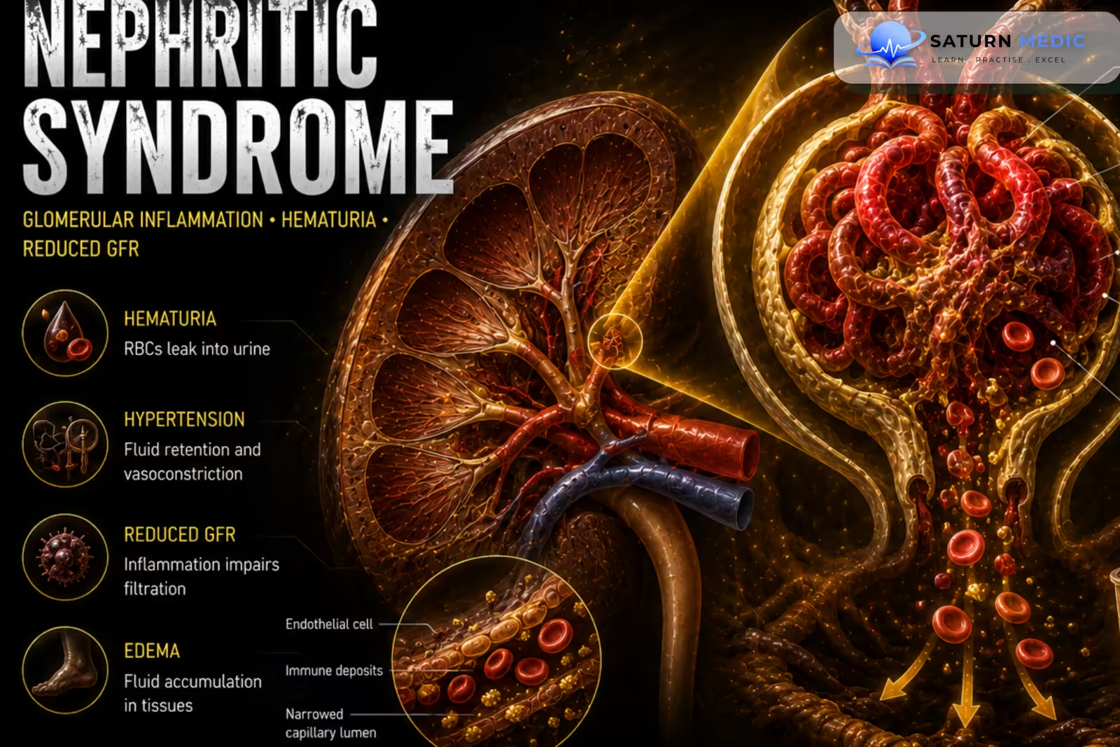
Nephritic syndrome is a clinical syndrome caused by glomerular inflammation, resulting in hematuria, reduced glomerular filtration, variable proteinuria, hypertension, and fluid retention
Also called
ICD-10
Specialty
Onset
Reviewed
-
OverviewOverview
-
Etiology & Risk FactorsEtiology & Risk Factors
-
PathophysiologyPathophysiology
-
Clinical PresentationClinical Presentation
-
History TakingHistory Taking
-
Physical ExaminationPhysical Examination
-
InvestigationsInvestigations
-
DiagnosisDiagnosis
-
ManagementManagement
-
ComplicationsComplications
-
PrognosisPrognosis
-
Key Points / Clinical PearlsKey Points / Clinical Pearls
Overview
–Nephritic syndrome results from inflammatory injury to the glomeruli, causing disruption of the glomerular filtration barrier and reduced glomerular filtration rate (GFR).
-Typical features include:
- Hematuria, often with RBC casts
- Mild–moderate proteinuria
- Hypertension
- Oliguria
- Edema
- Reduced GFR
Common causes include post-streptococcal glomerulonephritis, IgA nephropathy, rapidly progressive glomerulonephritis (RPGN), and systemic diseases such as systemic lupus erythematosus (SLE) and vasculitis.
Etiology & Risk Factors
-Etiology
–Nephritic syndrome is caused by glomerular inflammation, usually due to immune-mediated glomerular injury.
–Major causes include:
- Post-streptococcal glomerulonephritis
- IgA nephropathy
- IgA vasculitis
- Lupus nephritis
- ANCA-associated vasculitis
- Anti-glomerular basement membrane (anti-GBM) disease
- Membranoproliferative glomerulonephritis (MPGN)
-Risk Factors:
- Recent streptococcal infection
- Autoimmune disease, especially SLE
- Systemic vasculitis
- Recent infection
- Previous episodes of glomerulonephritis
Pathophysiology
Immune-mediated glomerular injury → glomerular inflammation → increased glomerular permeability + reduced GFR → RBC leakage into urine + oliguria → sodium and water retention → hypertension + edema
Clinical Presentation
-Symptoms:
- Hematuria
- Dark, tea-colored or cola-colored urine
- Reduced urine output
- Facial/periorbital swelling
- Peripheral edema
- Headache due to hypertension
- Fatigue
-Signs:
- Hypertension
- Periorbital edema
- Peripheral edema
- Reduced urine output
- Hematuria
- Signs of fluid overload
History Taking
-Ask about:
- Onset and duration of hematuria
- Urine color and volume
- Recent sore throat or skin infection
- Recent respiratory or gastrointestinal infection
- Edema and weight gain
- Headache or visual symptoms
- Symptoms of SLE
- Rash or joint pain
- Sinus or respiratory symptoms suggesting vasculitis
- Hemoptysis suggesting pulmonary-renal syndrome
Physical Examination
-General Examination
- Blood pressure
- Periorbital and peripheral edema
- Fluid overload
- Weight gain
- Signs of severe hypertension
-System-Specific Examination:
- Urinalysis findings
- Signs of pulmonary edema
- Skin rash or purpura
- Joint inflammation
- Sinus or respiratory abnormalities when vasculitis is suspected
Investigations
-Biochemistry / Specific Tests:
Serum creatinine and urea — assess kidney function
Electrolytes — assess renal complications
Urinalysis — hematuria and proteinuria
Urine microscopy — dysmorphic RBCs and RBC casts
Urine protein quantification — assess degree of proteinuria
Complement C3/C4
Antistreptolysin O (ASO) titer and/or anti-DNase B when post-streptococcal disease is suspected
Antinuclear antibody (ANA) and anti-double-stranded DNA (anti-dsDNA) when SLE is suspected
ANCA when vasculitis is suspected
Anti-GBM antibodies when anti-GBM disease is suspected
-Imaging
Renal ultrasound — assess kidney size and exclude obstruction when indicated
-Special / Confirmatory Tests
Kidney biopsy may be required when the cause is unclear, renal function is rapidly deteriorating, or RPGN is suspected.
Diagnosis
-Nephritic syndrome is diagnosed clinically by the combination of:
- Hematuria
- Dysmorphic RBCs and/or RBC casts
- Proteinuria, usually less than nephrotic-range
- Reduced GFR or AKI
- Hypertension and/or edema
-The underlying cause is determined using complement levels, serology, infection testing, and sometimes kidney biopsy.
Management
1. First-Line / Emergency Management
There is no routine emergency treatment.
If severe complications are present:
Control severe hypertension
Treat pulmonary edema/fluid overload
Manage hyperkalemia
Initiate dialysis when indicated
2. Definitive Treatment
Treat the underlying cause of glomerulonephritis.
Examples:
Post-streptococcal GN → supportive treatment
IgA nephropathy → supportive renal protection; immunosuppression in selected patients
Lupus nephritis → immunosuppressive therapy according to class/severity
ANCA-associated vasculitis → immunosuppressive therapy
Anti-GBM disease → urgent plasma exchange + immunosuppression
3. Medical Treatment
ACE inhibitors or ARBs — reduce proteinuria and control hypertension when appropriate
Diuretics — manage clinically significant edema/fluid overload
Corticosteroids — selected immune-mediated causes
Other immunosuppressive agents — according to the underlying disease
Antibiotics — when an active bacterial infection requires treatment
4. Surgical / Procedural Treatment
No routine surgical treatment.
Dialysis may be required for severe AKI or complications such as refractory hyperkalemia, pulmonary edema, severe acidosis, or uremia.
5. Supportive Management
Salt restriction
Fluid restriction when significant fluid overload or oliguria is present
Blood pressure control
Monitor renal function and electrolytes
Complications
- Acute kidney injury
- Severe hypertension
- Pulmonary edema
- Hyperkalemia
- Metabolic acidosis
- Uremia
- Chronic kidney disease
- End-stage kidney disease
Prognosis
-Prognosis depends mainly on the underlying cause, severity of renal impairment, and response to treatment.
-Post-infectious nephritic syndromes often recover well, particularly in children, while rapidly progressive or systemic immune-mediated disease can progress to chronic kidney disease or kidney failure.
Key Points / Clinical Pearls
- Nephritic syndrome is caused by glomerular inflammation.
- The classic finding is hematuria.
- Urine may appear tea-colored or cola-colored.
- Dysmorphic RBCs and RBC casts indicate glomerular hematuria.
- Proteinuria is usually less than nephrotic-range.
- Hypertension is common due to sodium and water retention.
- Reduced GFR may cause oliguria and AKI.
- Common causes include post-streptococcal GN and IgA nephropathy.
- SLE and systemic vasculitis are important systemic causes.
- Complement levels help narrow the underlying cause.
- Serological testing should be guided by the clinical presentation.
- Kidney biopsy may be required when the diagnosis or cause is unclear.
- Kidney Disease: Improving Global Outcomes (KDIGO) Glomerular Diseases Work Group. KDIGO 2021 Clinical Practice Guideline for the Management of Glomerular Diseases. Kidney Int. 2021;100(4S):S1-S276. KDIGO .
- Floege J, Amann K. Primary Glomerulonephritides. Lancet. 2016;387(10032):2036-2048. The Lancet .
- McGrogan A, Franssen CF, de Vries CS. The Incidence of Primary Glomerulonephritis Worldwide: A Systematic Review of the Literature. Nephrol Dial Transplant. 2011;26(2):414-430.
- Couser WG. Primary Membranous Nephropathy. Clin J Am Soc Nephrol. 2017;12(6):983-997. Clinical Journal of the American Society of Nephrology .
- National Institute of Diabetes and Digestive and Kidney Diseases (NIDDK). Glomerular Diseases . National Institutes of Health.
- National Library of Medicine (NIH). Glomerulonephritis . StatPearls.