

Clinical Subject Page
Abdominal aortic aneurysm (AAA)
ICD-10
Specialty
Onset
Reviewed
-
OverviewOverview
-
Etiology & Risk FactorsEtiology & Risk Factors
-
PathophysiologyPathophysiology
-
Clinical PresentationClinical Presentation
-
History TakingHistory Taking
-
Physical ExaminationPhysical Examination
-
InvestigationsInvestigations
-
DiagnosisDiagnosis
-
ManagementManagement
-
ComplicationsComplications
-
PrognosisPrognosis
-
Key Points / Clinical PearlsKey Points / Clinical Pearls
Overview
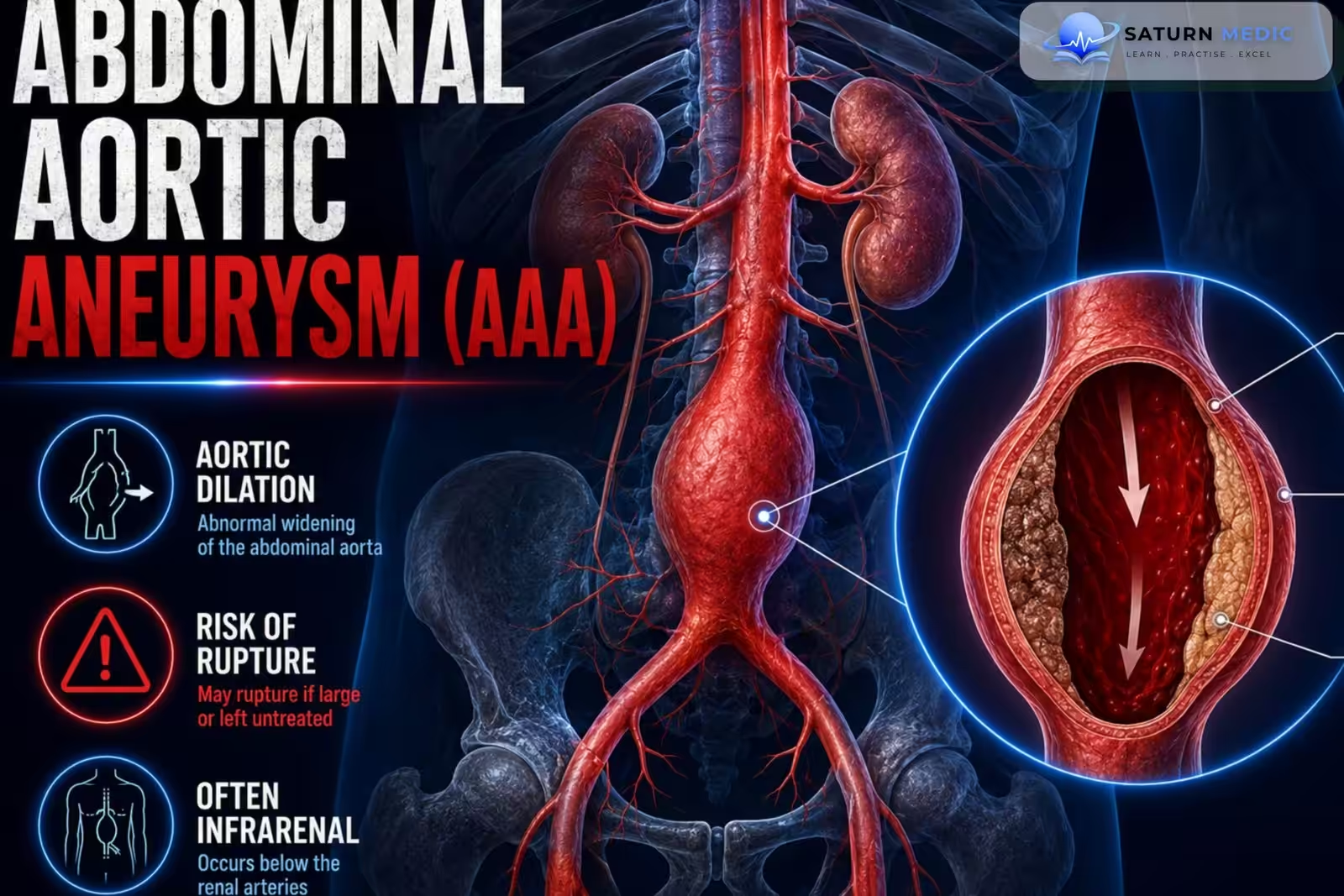
Abdominal aortic aneurysm (AAA) is a localized dilation of the abdominal aorta measuring ≥3.0 cm in diameter or ≥50% greater than the normal vessel diameter, resulting from weakening of the aortic wall and increasing the risk of rupture.
Etiology & Risk Factors
Etiology for Abdominal aortic aneurysm (AAA)
Atherosclerosis (most common)
Chronic hypertension
Smoking-related vascular degeneration
Connective tissue disorders (Marfan, Ehlers-Danlos)
Inflammatory aortitis
Infectious (mycotic) aneurysm
Previous aortic trauma
Risk Factors for Abdominal aortic aneurysm (AAA)
Age ≥65 years
Male sex
Smoking (strongest modifiable risk factor)
Hypertension
Family history of Abdominal aortic aneurysm (AAA)
Hyperlipidemia
Coronary artery disease
Peripheral arterial disease
Pathophysiology
- Inflammation and proteolytic degeneration of collagen, elastin, and smooth muscle cells weaken the abdominal aortic wall.
- Loss of wall strength leads to progressive widening (aneurysm).
- Mechanical stress (especially from hypertension) further enlarges the aneurysm and may cause rupture.
- The dilated vessel disrupts laminar blood flow, producing turbulent flow.
- Mural thrombi may develop within the aneurysm and cause peripheral thromboembolism.
Simple Flow
Wall degeneration → Weak abdominal aortic wall → Aneurysm formation → Turbulent blood flow → Thrombus formation ± Rupture
Clinical Presentation
Symptoms
Usually asymptomatic
Chest pain
Back pain
Hoarseness
Dysphagia
Dyspnea
Cough
Signs
Early diastolic murmur (aortic regurgitation)
Features of connective tissue disorders
Signs of compression of adjacent structures
Hypotension or shock (rupture)
History Taking
Key Questions
Sudden abdominal or back pain?
Pulsatile abdominal sensation?
History of smoking?
Hypertension?
Family history of Abdominal aortic aneurysm (AAA)?
Previous aneurysm?
Syncope or dizziness?
Peripheral vascular disease?
Red Flags for Abdominal aortic aneurysm (AAA)
Sudden severe abdominal or back pain
Hypotension
Syncope
Pulsatile abdominal mass
Signs of hemorrhagic shock
Physical Examination
Inspection
May show a visible pulsation in thin patients
Palpation
Pulsatile abdominal mass (classic finding)
Auscultation
Abdominal bruit
General Examination
Assess peripheral pulses
Check for signs of hypovolemic shock if rupture is suspected
If Rupture of Abdominal aortic aneurysm (AAA) Occurs
Hypotension
Tachycardia
Severe abdominal or back pain
Signs of shock
Investigations
Laboratory
CBC
Renal function
Coagulation profile
Blood type & crossmatch (if rupture suspected)
Imaging
Abdominal ultrasound (screening & surveillance)
CT angiography (gold standard for diagnosis and operative planning)
Additional Tests
ECG (exclude cardiac causes)
Chest X-ray (if indicated)
Diagnosis
Abdominal Aortic Aneurysm (AAA) · Diagnostic Criteria
| Category | Diameter | Action |
|---|---|---|
| Normal infrarenal aorta | <3.0 cm | No follow-up required |
| Small AAA | 3.0–4.4 cm | Annual ultrasound surveillance; address cardiovascular risk factors |
| Medium AAA | 4.5–5.4 cm | Ultrasound surveillance every 3 months; vascular surgery referral |
| Large AAA | ≥5.5 cm | Elective surgical repair indicated (open or EVAR) regardless of symptoms |
| Rapid expansion | ≥1 cm/year or ≥0.5 cm in 6 months | Treat as high risk — expedite surgical referral regardless of absolute size |
| Symptomatic AAA (pain, tenderness) | Any size | Urgent CT angiography and surgical referral — treat as impending rupture |
| Ruptured AAA | Any size | Surgical emergency — classic triad: abdominal/back pain, pulsatile mass, hypotension |
Management
Medical Management
Smoking cessation
Blood pressure control
Statin therapy
Antiplatelet therapy (if indicated)
Regular imaging surveillance for small aneurysms
Surgical Management
Endovascular aneurysm repair (EVAR)
Open surgical repair
Immediate repair for ruptured Abdominal aortic aneurysm (AAA)
Elective repair for symptomatic or large aneurysms (typically ≥5.5 cm in men, or rapidly expanding aneurysms)
Complications
- Aneurysm rupture
- Massive hemorrhage
- Thromboembolism
- Distal limb ischemia
- Aortic dissection (rare)
- Death
Prognosis
- Small Abdominal aortic aneurysm (AAA) have a good prognosis with surveillance.
- Risk of rupture increases markedly with aneurysm size and expansion rate.
- Ruptured Abdominal aortic aneurysm (AAA) has a very high mortality despite emergency surgery.
Key Points / Clinical Pearls
- Abdominal Aortic Aneurysm (AAA) is a permanent dilatation of the abdominal aorta, commonly defined as an aortic diameter ≥3.0 cm.
- Most AAAs are asymptomatic and are discovered incidentally or through screening.
- The strongest risk factors are smoking, increasing age, male sex, and family history of AAA.
- Other important risk factors include hypertension, hyperlipidemia, and atherosclerotic cardiovascular disease.
- A symptomatic AAA may cause abdominal, back, or flank pain.
- A pulsatile abdominal mass may be present but is not always detectable.
- AAA rupture causes sudden severe abdominal or back pain and may produce hypotension, syncope, or shock; it is a life-threatening emergency.
- Abdominal ultrasound is the preferred test for screening and is highly accurate for detecting AAA.
- CT angiography is particularly important for symptomatic patients and for preoperative anatomical assessment.
- Small asymptomatic AAAs are generally managed with surveillance and cardiovascular risk-factor control rather than immediate repair.
- Smoking cessation and management of cardiovascular risk factors are essential in patients with AAA.
- National Center for Biotechnology Information (NIH). Abdominal Aortic Aneurysm, StatPearls.
- US Preventive Services Task Force. Screening for Abdominal Aortic Aneurysm: US Preventive Services Task Force Recommendation Statement. JAMA. 2019;322:2211-2218. PMID: 31821437.
- Isselbacher EM, Preventza O, Hamilton Black J 3rd, et al. 2022 ACC/AHA Guideline for the Diagnosis and Management of Aortic Disease. Circulation. 2022;146:e334-e482. PMID: 36334952.
- MedlinePlus, National Library of Medicine (NIH). Abdominal Aortic Aneurysm: Medical Encyclopedia.
- National Center for Biotechnology Information (NIH). Ruptured Abdominal Aortic Aneurysm, StatPearls.