

Clinical Subject Page
Aortic Stenosis
Also called
ICD-10
Specialty
Onset
Reviewed
-
OverviewOverview
-
Etiology & Risk FactorsEtiology & Risk Factors
-
PathophysiologyPathophysiology
-
Clinical PresentationClinical Presentation
-
History TakingHistory Taking
-
Aortic Stenosis Vs Aortic RegurgitationAortic Stenosis Vs Aortic Regurgitation
-
InvestigationsInvestigations
-
DiagnosisDiagnosis
-
ManagementManagement
-
ComplicationsComplications
-
PrognosisPrognosis
-
Key Points / Clinical PearlsKey Points / Clinical Pearls
Overview
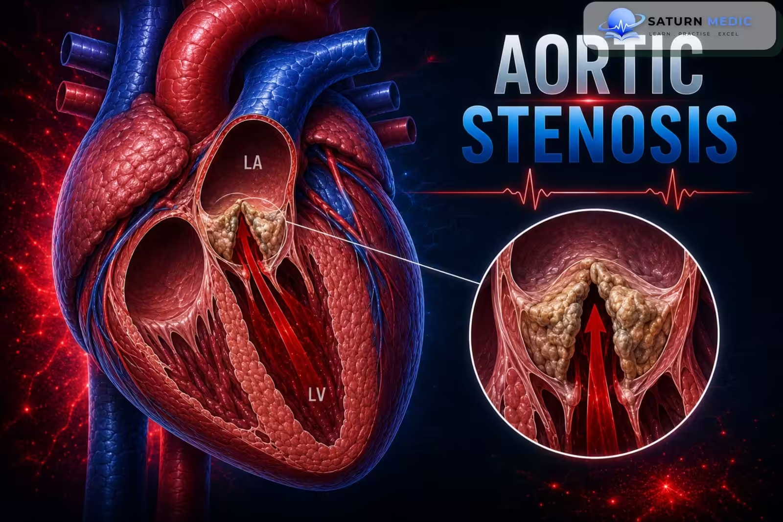
Aortic stenosis (AS), commonly known as narrowing of the aortic valve, is a heart valve condition in which the aortic valve becomes narrowed, making it harder for blood to flow from the left ventricle into the aorta. The condition often progresses slowly and may not cause symptoms for years, but it can eventually lead to shortness of breath, chest pain (angina), or fainting (syncope).
Etiology & Risk Factors
Etiology of Aortic Stenosis
The most common causes include:
- Aortic valve sclerosis (calcification and fibrosis of the aortic valve leaflets) – most common cause
- Bicuspid aortic valve (BAV) – a congenital abnormality that predisposes the valve to early calcification and degeneration
- Rheumatic fever – a less common cause in high-income countries but remains important in lower-income countries
Risk Factors
- Advanced age (risk increases with aging)
- Bicuspid aortic valve (congenital)
- Risk factors for atherosclerosis, as aortic valve sclerosis has a similar pathophysiology to atherosclerosis
Pathophysiology
- Narrowing of the aortic valve during systole obstructs blood flow from the left ventricle (LV) into the aorta.
- This obstruction causes increased pressure in the LV, leading to concentric left ventricular hypertrophy (thickening of the LV wall).
- The hypertrophied LV results in:
- Increased myocardial oxygen demand
- Impaired ventricular filling during diastole, which can lead to left-sided heart failure
- Reduced coronary flow reserve
- Initially, cardiac output is maintained through compensatory mechanisms.
- As the disease progresses, the LV becomes less compliant, cardiac output decreases, and pressure backs up into the pulmonary veins and capillaries, increasing the workload on the right heart and potentially leading to right-sided heart failure.
-Flow summary:
Aortic valve narrowing → LV outflow obstruction → ↑ LV pressure → Concentric LV hypertrophy → ↓ Diastolic filling & ↑ O₂ demand → ↓ Cardiac output → Left heart failure → Pulmonary congestion → Right heart failure (late).
Clinical Presentation
Common symptoms (remember: “SAD”)
- Syncope (dizziness or fainting)
- Angina (chest pain)
- Dyspnea (usually on exertion)
Physical examination
- Weak and delayed carotid pulse (pulsus parvus et tardus)
- Decreased pulse pressure (small blood pressure amplitude)
- Palpable systolic thrill over the carotids and aorta
Auscultation findings
- Harsh crescendo-decrescendo systolic ejection murmur
- Best heard at the 2nd right intercostal space
- Murmur radiates to the carotid arteries
- Soft S2
- S4 heart sound (due to decreased LV compliance)
- Early systolic ejection click
History Taking
- Do you get short of breath when walking or climbing stairs?”
- “Have you had any chest pain or tightness, especially during activity?”
- “Have you ever felt dizzy or fainted?”
- “When did these symptoms start?”
- “Do your symptoms get worse with exercise?”
- “Have your symptoms become more frequent or severe?”
- “Have you ever been told you have a heart murmur or a heart valve problem?”
- “Do you have a history of rheumatic fever or congenital heart disease?”
- “Has anyone in your family had heart valve disease?”
Aortic Stenosis Vs Aortic Regurgitation
Valvular Heart Disease · Aortic Stenosis vs Aortic Regurgitation
| Parameter | Aortic Stenosis | Aortic Regurgitation |
|---|---|---|
| Pathology | Narrowed aortic valve orifice → LV must generate high pressure to eject blood (pressure overload) | Incompetent aortic valve → blood regurgitates back into LV during diastole (volume overload) |
| LV Response | Concentric hypertrophy — wall thickens, cavity size normal or reduced | Eccentric hypertrophy — wall thickens AND cavity dilates (Laplace law) |
| Onset | Usually chronic & insidious (years of compensated disease before symptoms) | Can be chronic (insidious) or acute (endocarditis, aortic dissection — surgical emergency) |
| Classic Triad / Symptoms |
Syncope Angina Dyspnea
SAD triad — appear in this order as severity progresses. Syncope on exertion is a red flag.
|
Exertional dyspnea Palpitations Angina
Long asymptomatic phase. Symptoms indicate LV decompensation. Acute AR: flash pulmonary edema.
|
| Pulse | Pulsus parvus et tardus — small volume, slow-rising, delayed peak | Corrigan's (water-hammer) pulse — large volume, bounding, rapid rise and collapse |
| Blood Pressure | Narrow pulse pressure (<40 mmHg); systolic BP may be low in severe AS | Wide pulse pressure (>60–80 mmHg); high systolic, low diastolic |
| Apex Beat | Sustained (heaving), non-displaced — pressure-loaded LV | Displaced laterally & downward, hyperdynamic — volume-loaded dilated LV |
| Murmur Type | Ejection systolic (crescendo-decrescendo) — heard best at aortic area; radiates to carotids | Early diastolic decrescendo — heard best at left sternal border, leaning forward in expiration |
| Additional Murmurs | S4 (stiff LV); paradoxically split S2 (severe); ejection click (bicuspid valve) | Austin Flint murmur (low-pitched mid-diastolic rumble at apex); S3 (dilated LV) |
| ECG Findings | LVH with strain pattern (ST depression & T-wave inversion in V5–V6, I, aVL); LV strain | LVH (voltage criteria); broad notched P (if LA enlarges); LV volume overload pattern |
| CXR Findings | Normal heart size (concentric LVH); calcified aortic valve; post-stenotic aortic dilatation | Cardiomegaly (LV enlargement); prominent aorta; pulmonary edema if decompensated |
| Echo / Severity | Valve area: mild >1.5 cm², moderate 1.0–1.5 cm², severe <1.0 cm²; mean gradient severe >40 mmHg | Regurgitant fraction: mild <30%, moderate 30–49%, severe ≥50%; LV end-systolic diameter |
| Medical Management | No effective medical therapy to slow progression. Manage comorbidities (HTN, AF). Avoid vasodilators in severe AS. | Vasodilators (nifedipine, ACEi/ARB) to reduce afterload & preload in chronic AR. Bridge to surgery. |
| Surgical Indications | Severe AS + symptoms; severe AS + EF <50%; severe AS undergoing other cardiac surgery | Severe AR + symptoms; severe AR + EF <50%; severe AR + LV ESD >50 mm |
| Intervention Options | SAVR TAVR Balloon valvuloplasty (palliation only) | SAVR Valve repair (if feasible) — TAVR emerging but less established |
| Prognosis | Untreated symptomatic severe AS: 50% mortality at 2–3 yrs. Rapid decline after symptom onset. | Chronic: long compensated phase. Once symptomatic: 10–20% annual mortality without surgery. |
Radiation: Both carotids — key distinguishing feature
Character: Harsh, rasping; peaks in mid-systole; later peak = more severe
Maneuvers: Louder on squatting (increased preload); softer on standing/Valsalva
S2: Soft or absent A2; paradoxical splitting in severe AS
Extras: Ejection click (bicuspid valve); S4 (non-compliant LV)
Position: Patient leaning forward, end-expiration — brings aorta closer to chest wall
Character: Blowing, high-pitched; begins immediately after S2
Maneuvers: Louder on squatting & handgrip (increased afterload)
Austin Flint murmur: Low-pitched mid-diastolic rumble at apex — regurgitant jet impinges on anterior mitral leaflet
Extras: S3 (dilated LV); wide pulse pressure
Investigations
- Transthoracic echocardiography (TTE) – Gold standard for diagnosis; assesses valve structure, severity of stenosis, pressure gradient, and left ventricular function.
- Transesophageal echocardiography (TEE) – Used if TTE is inconclusive or for operative planning.
- ECG – May show left ventricular hypertrophy (LVH) and nonspecific ST-T wave changes.
- Chest X-ray – May show aortic valve calcification, LV enlargement, pulmonary congestion, or poststenotic dilation of the aorta.
- Laboratory tests – BNP/NT-proBNP and troponin may be helpful but are not routinely diagnostic.
- Cardiac catheterization/coronary angiography – Used when noninvasive tests are inconclusive or before valve intervention in selected patients.
- Cardiac CT or MRI – Used in selected patients for further evaluation and preoperative planning.
Diagnosis
Aortic Stenosis · Diagnostic Criteria & Severity Classification
Severe AS threshold
Severe AS threshold
Severe AS threshold
| Modality | Finding | Notes |
|---|---|---|
| History |
Angina
Syncope
Dyspnea
SAD triad — mean survival: angina 5 yrs, syncope 3 yrs, dyspnea (HF) 1–2 yrs
|
Symptoms appear late. Long asymptomatic compensated phase. Symptom onset = inflection point for prognosis. |
| Pulse | Pulsus parvus et tardus — small volume, slow-rising, delayed peak | Best assessed at carotid. Most specific clinical sign of severe AS. May be absent if coexisting AR or hypertension. |
| Auscultation |
Harsh ejection systolic murmur — crescendo-decrescendo, peaks in mid-to-late systole
Best heard: Right 2nd ICS, radiates to carotids
Gallavardin phenomenon: high-frequency component radiates to apex, mimics MR
|
Later systolic peak = more severe. Soft or absent A2 (calcified valve). S4 common. Ejection click in bicuspid valve (before calcification). |
| Maneuvers | Louder: squatting, leg raise Softer: standing, Valsalva | Contrast with HCM: AS murmur decreases on standing; HCM murmur increases. Valsalva differentiates AS from HCM. |
| ECG |
LVH (Sokolow-Lyon: S in V1 + R in V5/V6 ≥35 mm)
LV strain pattern — ST depression & T-wave inversion in V5–V6, I, aVL
LBBB, AF (late features)
|
ECG may be normal even with severe AS. Not used for severity grading. AF carries worse prognosis — loss of atrial kick critical in stiff LV. |
| Chest X-Ray |
Calcified aortic valve (best seen on lateral CXR or fluoroscopy)
Post-stenotic aortic dilatation — dilated proximal ascending aorta
Heart size normal (concentric LVH) unless decompensated; then cardiomegaly + pulmonary edema
|
Absence of valve calcification on echo/CT in a patient >65 yrs makes severe AS unlikely. Calcium load correlates with severity. |
| Echocardiography |
Transthoracic Echo (TTE) — First-line imaging modality TOE / TEE — if TTE suboptimal 3D Echo — planimetry of valve area |
TTE is the gold standard for diagnosis and severity grading. Provides valve morphology, gradient, valve area, LV function, and other valve disease. |
| CT / CMR |
CT Aortic Calcium Score (ACS): ≥2000 AU (men) / ≥1200 AU (women) = severe AS
CT Planimetry: direct anatomical valve area measurement
CMR: LV mass, fibrosis (late gadolinium enhancement), flow assessment
|
CT calcium scoring used when echo is discordant. CMR useful for detecting myocardial fibrosis — marker of irreversible damage and poor post-op outcome. |
| Cardiac Catheterization |
Gorlin formula: AVA = CO / (SEP × HR × 44.3 × √ΔP)
Direct measurement of transvalvular gradient
|
Rarely needed solely for AS diagnosis. Used when echo is non-diagnostic or discordant. Coronary angiography before surgery (if ≥40 yrs or CAD risk factors). |
| Exercise Testing | Symptom-limited treadmill/bicycle stress echo — used in asymptomatic severe AS | Contraindicated in symptomatic severe AS. Uncovers occult symptoms, abnormal BP response, and exercise-induced LV dysfunction — guides intervention timing. |
• True severe AS: AVA remains <1.0 cm², gradient increases ≥40 mmHg
• Pseudo-severe AS: AVA normalises >1.0 cm² as flow increases
Use CT calcium scoring (AU) and AVAi to confirm severity. Often missed and undertreated.
• 3D echo or CT planimetry for true AVA
• CT calcium score
• Velocity ratio (flow-independent)
True moderate AS is also in the differential.
Management
Management of Aortic Stenosis
- Mild or asymptomatic disease
- Conservative management with regular monitoring (serial echocardiography).
- Treat comorbidities (e.g., hypertension, diabetes).
- Symptomatic or severe aortic stenosis
- Aortic valve replacement (AVR) is the definitive treatment.
- Surgical AVR (SAVR): Typically for younger patients with low to moderate surgical risk.
- Transcatheter AVR (TAVR): Typically for older patients or those at high surgical risk.
- Acute complications
- Immediate cardiology/cardiothoracic surgery consultation.
- Stabilize the patient while expediting definitive valve replacement.
Complications
Complications of Aortic Stenosis
Left ventricular failure
Acute heart failure
Cardiogenic shock
Atrial fibrillation with rapid ventricular response (may occur in severe disease)
Complications After Valve Replacement
Stroke/thromboembolism
Major bleeding
Renal failure
Arrhythmias (e.g., atrial fibrillation, atrioventricular block)
Aortic regurgitation/paravalvular leak
Infective endocarditis
Prognosis
- Asymptomatic patients: Annual mortality is < 1%.
- Symptomatic patients: If left untreated, > 50% die within 2 years.
- The worst prognosis is seen in patients with heart failure symptoms, followed by syncope and angina.
Key Points / Clinical Pearls
- Chronic, progressive narrowing of the aortic valve.
- Most common cause: Age-related calcific aortic valve sclerosis.
- Classic symptom triad (SAD): Syncope, Angina, Dyspnea.
- Classic murmur: Harsh crescendo-decrescendo systolic murmur radiating to the carotids.
- Gold standard diagnosis: Transthoracic echocardiography (TTE).
- Definitive treatment: Aortic valve replacement (AVR) (SAVR or TAVR).
- Once symptoms develop, prompt evaluation for valve replacement is essential because prognosis worsens significantly without treatment.
- Pujari SH, Agasthi P. National Center for Biotechnology Information (NIH). Aortic Stenosis, StatPearls.
- Otto CM, Nishimura RA, Bonow RO, et al. 2020 ACC/AHA Guideline for the Management of Patients With Valvular Heart Disease. Circulation. 2021;143:e72-e227. PMID: 33972115.
- National Center for Biotechnology Information (NIH). Transcatheter Aortic Valve Replacement, StatPearls.
- Highlights From the 2020 ACC/AHA Guidelines on Valvular Heart Disease: Review of 2020 AHA/ACC VHD Guidelines. PMC9707478.
- MedlinePlus, National Library of Medicine (NIH). Aortic Stenosis: Medical Encyclopedia.