

Clinical Subject Page
Atrial fibrillation (AFib)
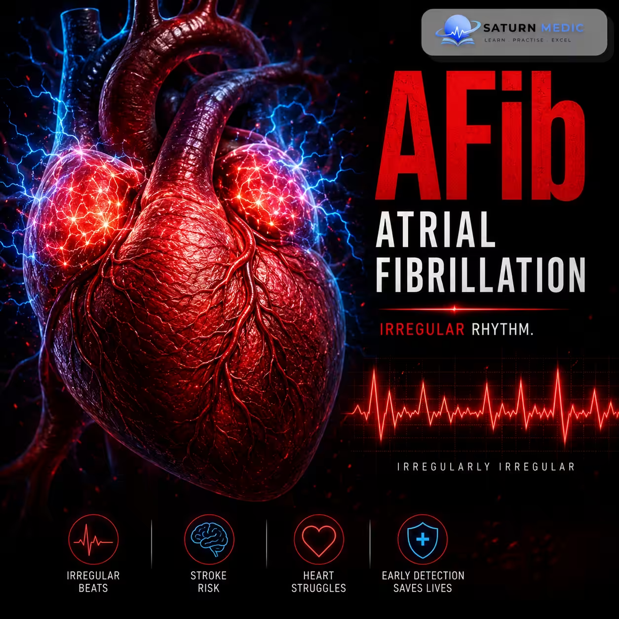
Atrial fibrillation (AFib) is a common type of abnormal heart rhythm in which the upper chambers of the heart (the atria) beat in a rapid, uncoordinated way. This causes the heartbeat to become irregular.
ICD-10
Specialty
Onset
Reviewed
-
OverviewOverview
-
Etiology & Risk FactorsEtiology & Risk Factors
-
PathophysiologyPathophysiology
-
Clinical PresentationClinical Presentation
-
History TakingHistory Taking
-
Physical ExaminationPhysical Examination
-
InvestigationsInvestigations
-
DiagnosisDiagnosis
-
ManagementManagement
-
ComplicationsComplications
-
PrognosisPrognosis
-
Key Points / Clinical PearlsKey Points / Clinical Pearls
Overview
Atrial fibrillation (AFib) is the most common sustained abnormal heart rhythm. In AFib, the upper chambers of the heart (atria) beat very rapidly and irregularly, causing the heart rhythm to become irregularly irregular.
Etiology & Risk Factors
The exact cause of atrial fibrillation (AFib) is unknown. However, several underlying conditions and reversible triggers are associated with its development.
Reversible causes for Atrial fibrillation (AFib)
- Hyperthyroidism (thyrotoxicosis)
- Electrolyte imbalances
- Cardiothoracic surgery
- Myocarditis
- Pericarditis
- Myocardial infarction
- Alcohol use
- Excess caffeine
- Fever
- Recreational or pharmacologic drug use
- Pulmonary embolism
- Other triggers of tachycardia (e.g., pain, hypovolemia, anemia)
-Approximately 15% of people with AFib have no identifiable risk factors (idiopathic or “lone” AFib).
Risk Factors ;
- Advanced age
- Hypertension
- Diabetes mellitus
- Obesity
- Obstructive sleep apnea (OSA)
- Coronary artery disease (CAD)
- Valvular heart disease (especially mitral valve disease)
- Heart failure
- Hyperthyroidism
- Chronic lung disease (e.g., COPD)
- Excessive alcohol use (“holiday heart syndrome”)
Pathophysiology
Pathophysiology of Atrial Fibrillation (AFib)
- AFib is a supraventricular arrhythmia with rapid, disorganized electrical activity in the atria.
- The exact mechanism is not fully understood, but contributing factors include:
- Atrial dilation or hypertrophy due to volume overload or hemodynamic stress
- Atrial ischemia
- Inflammation of the atrial myocardium
- Abnormal ion conduction in atrial tissue
Mechanism
- Atrial fibrillation (AFib) is triggered by:
- Rapid electrical impulses from automatic foci near the pulmonary vein ostia (left atrium) or fibrotic atrial tissue.
- Less commonly, pre-excitation through an accessory pathway (e.g., WPW syndrome).
- Atrial fibrillation (AFib) is maintained by:
- Re-entry circuits and/or rapid ectopic firing.
- Enlarged or diseased atria make these abnormal circuits more likely.
- Atrial remodeling:
- Electrical changes occur within hours.
- Persistent AFib leads to atrial fibrosis and dilation over months.
- These changes make AFib more likely to continue (“AFib begets AFib”).
Effects
- Rapid, ineffective atrial contraction → blood stasis in the atria → thrombus formation → increased risk of stroke and systemic thromboembolism.
- Irregular conduction through the AV node → irregular ventricular rhythm and often tachycardia.
Clinical Presentation
Symptoms (stable Atrial fibrillation AFib)
Often asymptomatic
Palpitations
Fatigue
Dyspnea (shortness of breath)
Lightheadedness
Syncope
Signs of underlying heart disease (e.g., murmur from mitral stenosis)
Features of Unstable Atrial fibrillation (AFib)
Ischemic chest pain
Altered mental status
Pulmonary edema
Acute heart failure
Cardiogenic shock
Complications at Presentation
Stroke or TIA (most important)
Acute left heart failure with pulmonary edema
Other thromboembolic events (e.g., renal, splenic, intestinal, or limb infarction)
Tachycardia-induced cardiomyopathy
History Taking
- Palpitations
- Shortness of breath
- Chest pain
- Dizziness or lightheadedness
- Syncope (fainting)
- Fatigue
- Onset and duration of symptoms
- Previous episodes of AFib
- History of heart disease, hypertension, thyroid disease, or sleep apnea
- Recent illness, surgery, alcohol/caffeine intake, or stimulant/drug use
- Current medications (especially anticoagulants and rate/rhythm-control drugs)
-Red Flags for Atrial fibrillation (AFib)
- Chest pain (possible myocardial ischemia)
- Syncope
- Altered mental status
- Severe shortness of breath or pulmonary edema
- Signs of acute heart failure
- Cardiogenic shock or hemodynamic instability
- Symptoms of stroke/TIA (sudden weakness, facial droop, speech difficulty)
Physical Examination
- Vital signs
- Heart rate (tachycardia or, rarely, bradycardia)
- Blood pressure
- Oxygen saturation
- Pulse
- Irregularly irregular pulse (hallmark finding)
- Apical pulse deficit
- Cardiovascular examination
- Irregular heart rhythm on auscultation
- Murmurs suggesting underlying valvular disease (especially mitral valve disease)
- Respiratory examination
- Signs of pulmonary edema (crackles)
- Neurologic examination
- Assess for focal neurological deficits suggestive of stroke or TIA
- Assess for heart failure
- Peripheral edema
- Signs of acute heart failure or cardiogenic shock
Investigations
12-lead ECG (first-line)
Irregularly irregular rhythm
No discernible P waves
Cardiac rhythm monitoring (Holter/event monitor) if paroxysmal AFib is suspected and ECG is nondiagnostic
Blood tests
CBC – anemia, infection
Electrolytes – Na⁺, K⁺, Mg²⁺, Ca²⁺
Renal and liver function tests
Thyroid function tests (TFTs) – screen for hyperthyroidism
Cardiac imaging
Transthoracic echocardiogram (TTE)
Assess cardiac function
Detect structural heart disease (e.g., mitral valve disease)
Assess atrial enlargement
Transesophageal echocardiogram (TEE) (before cardioversion in selected patients)
Detect left atrial thrombus
Assess safety of rhythm control
Additional investigations (when indicated)
Chest X-ray (evaluate cardiopulmonary disease)
CT pulmonary angiography if pulmonary embolism is suspected
Diagnosis
Atrial Fibrillation · Diagnostic Criteria
| Pattern | Duration / definition | Spontaneous termination | Management implication |
|---|---|---|---|
| First detected | First diagnosed episode regardless of symptoms or duration | — | Establish rhythm; anticoagulate; investigate cause |
| Paroxysmal | Self-terminating, usually <48 h (up to 7 days) | Yes — within 7 days | Rate or rhythm control; CHA₂DS₂-VASc for OAC |
| Persistent | Lasts >7 days or requires cardioversion to terminate | No | Rhythm control preferred; anticoagulate ≥4 weeks before cardioversion |
| Long-standing persistent | Continuous AF >12 months; rhythm control still attempted | No | Consider ablation; strict anticoagulation |
| Permanent | AF accepted by patient and physician; no rhythm control attempted | No | Rate control only; lifelong anticoagulation |
Management of Atrial fibrillation (AFib)
1. Assess hemodynamic stability
Unstable AFib (e.g., hypotension, shock, acute heart failure, pulmonary edema, ischemic chest pain, altered mental status)
Immediate synchronized electrical cardioversion
Start anticoagulation if indicated, but do not delay emergency cardioversion
Stable AFib
Confirm diagnosis with ECG
Evaluate for structural heart disease (TTE)
Identify and treat reversible causes
2. Rate control
Goal: Control ventricular rate and relieve symptoms
First-line:
Beta-blockers
Nondihydropyridine calcium channel blockers
3. Rhythm control
Restore and maintain sinus rhythm with:
Electrical cardioversion
Antiarrhythmic drugs (e.g., flecainide, propafenone, amiodarone)
Catheter ablation
4. Treat underlying causes
Correct reversible triggers (e.g., hyperthyroidism, electrolyte abnormalities, alcohol use)
Manage cardiovascular risk factors and comorbidities
5. Anticoagulation : As Follows:
Atrial Fibrillation · CHA₂DS₂-VASc Score
| Letter | Risk factor | Definition / threshold | Points |
|---|---|---|---|
C |
Congestive Heart Failure | Signs/symptoms of HF or reduced LVEF (<40%) — includes HFpEF if symptomatic | +1 |
H |
Hypertension | Resting BP >140/90 mmHg on ≥2 occasions, or on antihypertensive therapy | +1 |
A₂ |
Age ≥ 75 years | High-risk age threshold — counts double due to markedly increased stroke risk | +2 |
D |
Diabetes mellitus | Fasting glucose >7 mmol/L (>125 mg/dL) or on oral hypoglycaemic / insulin therapy | +1 |
S₂ |
Stroke / TIA / thromboembolism | Prior ischemic stroke, TIA, or systemic thromboembolism — strongest individual predictor | +2 |
V |
Vascular disease | Prior MI, peripheral artery disease, or aortic plaque on imaging | +1 |
A |
Age 65–74 years | Intermediate age risk — scores 1 point (age ≥75 scores 2 points separately) | +1 |
Sc |
Sex category (female) | Female sex — risk modifier only; does not trigger anticoagulation alone (score must be ≥2 in females, ≥1 in males from other factors) | +1 |
Apixaban Rivaroxaban Dabigatran Edoxaban
Complications of Atrial fibrillation (AFib)
- Thromboembolic events (most important)
- Stroke/TIA
- Renal infarction
- Splenic infarction
- Intestinal ischemia
- Acute limb ischemia
- Acute left heart failure → pulmonary edema
- Tachycardia-induced cardiomyopathy
Prognosis of Atrial fibrillation (AFib)
- Generally good with appropriate treatment, especially with good rate/rhythm control and anticoagulation when indicated.
- The major determinant of prognosis is the risk of thromboembolic complications, particularly ischemic stroke.
- Prognosis is influenced by:
- Underlying heart disease and comorbidities
- Adequate rate or rhythm control
- Adherence to anticoagulation and other therapies
- Correction of reversible risk factors
Key Points / Clinical Pearls of Atrial fibrillation (AFib)
- Most common sustained cardiac arrhythmia.
- ECG: Irregularly irregular rhythm with absent P waves.
- Classic physical finding: Irregularly irregular pulse.
- Many patients are asymptomatic; common symptoms are palpitations, dyspnea, fatigue, dizziness, and syncope.
- Greatest complication: Ischemic stroke due to atrial thrombus formation.
- Diagnosis: 12-lead ECG; echocardiography evaluates structural heart disease.
- Nesheiwat Z, Goyal A, Jagtap M. National Center for Biotechnology Information (NIH). Atrial Fibrillation, StatPearls.
- Joglar JA, Chung MK, Armbruster AL, et al. 2023 ACC/AHA/ACCP/HRS Guideline for the Diagnosis and Management of Atrial Fibrillation. Circulation. 2024;149:e1-e156. PMID: 38033089.
- Ludhwani D, Wieters JS. National Center for Biotechnology Information (NIH). Paroxysmal Atrial Fibrillation, StatPearls.
- MedlinePlus, National Library of Medicine (NIH). Atrial Fibrillation: Health Topic.
- Noubiap JJ, Tan JJ, Teraoka JT, et al. Minimum National Prevalence of Diagnosed Atrial Fibrillation Inferred From California Acute Care Facilities. J Am Coll Cardiol. 2024;84:1501-1508. doi: 10.1016/j.jacc.2024.07.014.