

Clinical Subject Page
Atrial Flutter
ICD-10
Specialty
Onset
Reviewed
-
OverviewOverview
-
Etiology & Risk FactorsEtiology & Risk Factors
-
PathophysiologyPathophysiology
-
Clinical PresentationClinical Presentation
-
History TakingHistory Taking
-
Atrial Flutter Vs Atrial FibrillationAtrial Flutter Vs Atrial Fibrillation
-
InvestigationsInvestigations
-
DiagnosisDiagnosis
-
ManagementManagement
-
ComplicationsComplications
-
PrognosisPrognosis
-
Key Points / Clinical PearlsKey Points / Clinical Pearls
Overview
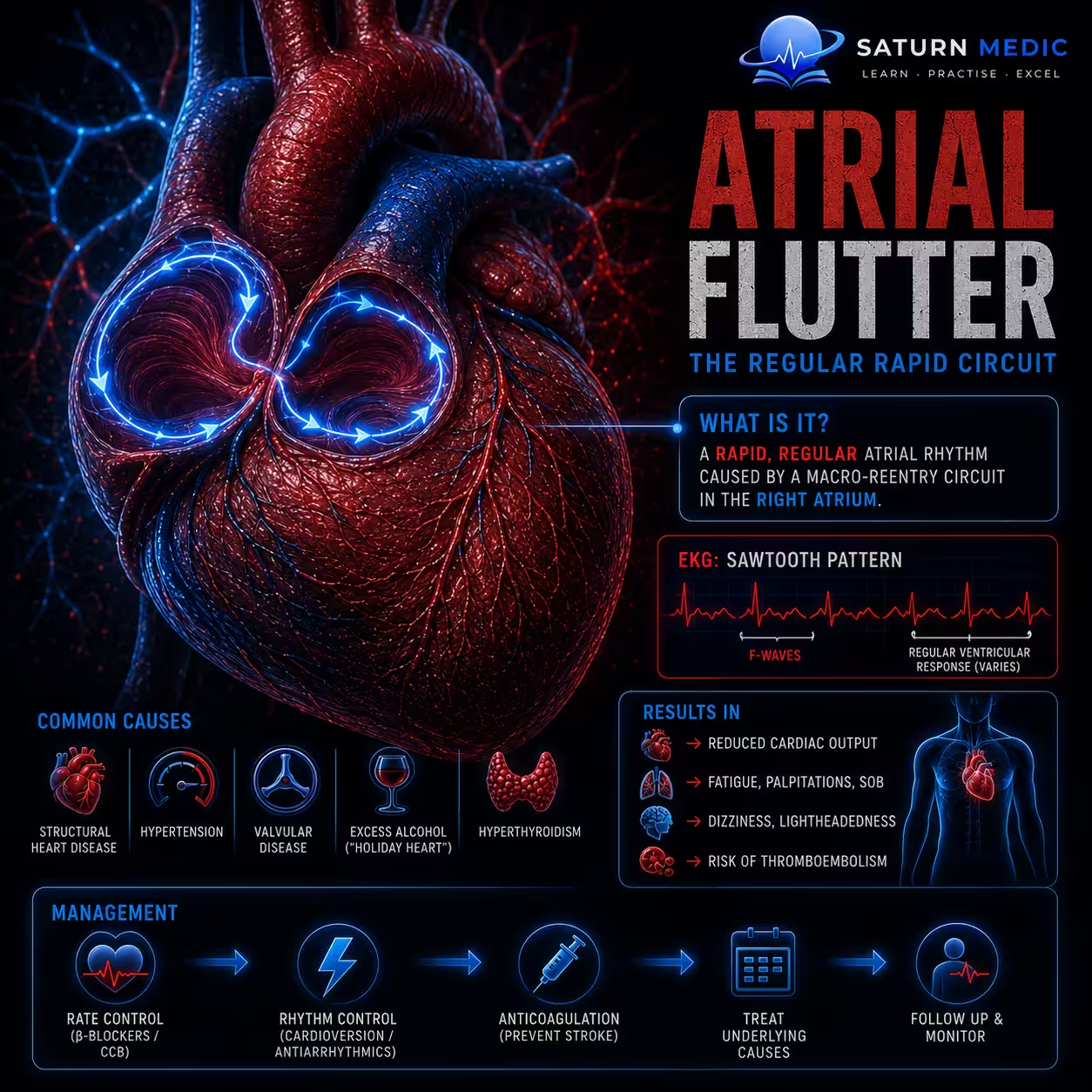
Atrial flutter is a supraventricular tachyarrhythmia (abnormally fast heart rhythm) caused by a macroreentrant electrical circuit within the atria. This rapid atrial activity causes the atria to beat very fast, while the ventricles usually beat at a regular but rapid rate.
Etiology & Risk Factors
Atrial flutter has causes similar to those of atrial fibrillation and is caused by a macroreentrant electrical circuit in the atria.
It may occur in patients with:
- Structural heart disease
- Valvular heart disease (especially mitral valve disease)
- Coronary artery disease
- Heart failure
- Cardiomyopathy
- Pericarditis
It may also occur after treatment of atrial fibrillation with:
- Flecainide
- Propafenone
- Amiodarone
Risk Factors
Cardiovascular
- Advanced age
- Hypertension
- Diabetes mellitus
- Smoking
- Obesity
- Obstructive sleep apnea (OSA)
Cardiac Disorders
- Coronary artery disease
- Valvular heart disease (especially mitral valve disease)
- Heart failure
- Cardiomyopathy
- Pericarditis
- Wolff-Parkinson-White (WPW) syndrome
- Sick sinus syndrome
Noncardiac Conditions
- COPD, pulmonary embolism, pneumonia
- Hyperthyroidism
- Electrolyte abnormalities (hypokalemia, hypomagnesemia)
- Sepsis or other causes of increased sympathetic activity
- Cocaine or amphetamine use
- Excess alcohol (“holiday heart syndrome”)
- Chronic kidney disease
Pathophysiology
- Atrial flutter is caused by a macroreentrant electrical circuit within the atria, resulting in rapid, organized atrial depolarization.
- The atria typically contract at a rate of about 300 beats/minute.
- The AV node blocks some impulses, so the ventricles usually beat more slowly (commonly with 2:1 or 4:1 AV conduction), producing a regular ventricular rhythm.
- There are two main types:
- Typical (Type I) atrial flutter: A reentrant circuit in the right atrium that travels around the tricuspid annulus through the cavotricuspid isthmus.
- Atypical (Type II) atrial flutter: Reentrant circuits that do not involve the cavotricuspid isthmus, often occurring in the left atrium or other atrial locations.
Simple Flow
Macroreentrant circuit in the atria → Rapid atrial rate (~300/min) → AV node blocks some impulses → Regular but rapid ventricular rate → Atrial flutter
Clinical Presentation
Common symptoms
- Palpitations
- Dizziness
- Fatigue
- Shortness of breath (dyspnea)
- Syncope (fainting)
Signs
- Tachycardia
- Regular pulse (in most patients)
- Signs of the underlying heart disease (e.g., murmur of mitral valve disease) may be present.
History Taking
- “Have you had a sore throat or strep throat in the past few weeks?”
- “Was it treated with antibiotics?”
- “Have you had a fever recently?”
- “Do you have pain or swelling that moves from one joint to another?”
- “Do you get short of breath during activity or when lying flat?”
- “Do you have chest pain or palpitations?”
- “Do you feel unusually tired or weak?”
- “Have you noticed any skin rash or painless lumps under your skin?”
- “Have you had any unusual jerky or involuntary movements?”
- “Have you ever been told you have a heart murmur, rheumatic fever, or a heart valve problem?”
- “Have you had rheumatic fever or repeated throat infections in the past?”
Atrial Flutter Vs Atrial Fibrillation
Atrial Flutter vs Atrial Fibrillation · Clinical Comparison
Investigations
- 12-lead ECG – Gold standard for diagnosis of Atrial Flutter.
- Classic finding: Sawtooth (F) waves (best seen in leads II, III, and aVF)
- Narrow QRS complexes
- Atrial rate around 300 beats/min
- Ventricular rate usually 75–150 beats/min depending on AV conduction.
- Cardiac rhythm monitoring
- Holter monitor or event recorder if episodes are intermittent.
- Continuous telemetry for hospitalized patients.
- Transthoracic echocardiography (TTE)
- Assesses cardiac function and looks for structural heart disease (e.g., valvular disease, atrial enlargement).
- Transesophageal echocardiography (TEE)
- Performed before cardioversion in selected patients to exclude left atrial thrombus.
- Laboratory tests
- CBC
- Electrolytes (Na⁺, K⁺, Mg²⁺, Ca²⁺)
- Thyroid function tests (TFTs)
- Renal and liver function tests
- Chest imaging
- Chest X-ray (CXR) if underlying cardiopulmonary disease is suspected.
- 12-lead ECG – Gold standard for diagnosis of Atrial Flutter.
Diagnosis
The diagnosis of atrial flutter is confirmed by a 12-lead ECG.
Diagnostic ECG findings:
- Sawtooth flutter (F) waves (best seen in leads II, III, and aVF) – hallmark finding
- Atrial rate ≈ 300 beats/min
- Ventricular rate usually 75–150 beats/min (depends on AV conduction, e.g., 2:1 or 4:1 block)
- Narrow QRS complexes (if there is no aberrant conduction)
- Usually a regular rhythm with fixed AV conduction, although variable conduction patterns can occur.
If episodes are intermittent or unclear:
- Holter monitor or other cardiac rhythm monitoring
- Electrophysiology (EP) study may be used to distinguish typical from atypical atrial flutter.
Management of Atrial Flutter
1. Initial Assessment
Assess hemodynamic stability (ABCDE approach).
Continuous cardiac monitoring and IV access.
Identify and treat reversible causes (e.g., electrolyte imbalance, hyperthyroidism).
2. Unstable Patient
Immediate synchronized electrical cardioversion (50–100 J biphasic).
Start anticoagulation as soon as indicated, but do not delay emergency cardioversion.
3. Stable Patient
Rate control
Control the ventricular rate with AV nodal blocking therapy (management is similar to atrial fibrillation).
Rhythm control
Electrical or pharmacologic cardioversion may be considered.
Catheter ablation is often the most effective treatment, with better success and lower recurrence than atrial fibrillation.
4. Anticoagulation
Assess stroke risk (CHA₂DS₂-VASc) and bleeding risk (HAS-BLED).
Anticoagulation recommendations are the same as for atrial fibrillation.
5. Long-Term Care
Regular cardiology follow-up.
Treat underlying cardiovascular disease and risk factors.
Encourage lifestyle modifications (e.g., weight loss, management of obstructive sleep apnea, reduction of alcohol intake).
Complications of Atrial Flutter
- Thromboembolism
- Ischemic stroke
- Systemic embolism
- Heart failure
- Tachycardia-induced cardiomyopathy
- Hypotension
- Myocardial ischemia
- Reduced cardiac output
- Worsening angina
- Syncope or presyncope
Prognosis of Atrial Flutter
- Generally good with appropriate treatment.
- Catheter ablation has better success and lower recurrence than in atrial fibrillation.
- Many patients respond well to rhythm control, but atrial flutter frequently progresses to atrial fibrillation, so long-term follow-up is important.
Key Points / Clinical Pearls of Atrial Flutter
- ECG hallmark of Atrial Flutter: Sawtooth (F) waves
- Atrial rate: ~300 beats/min
- Ventricular rhythm: Usually regular
- Unstable patient: Immediate synchronized electrical cardioversion
- Stable patient: Rate control or rhythm control
- Catheter ablation is often the most effective long-term treatment
- Assess need for anticoagulation using the same recommendations as for atrial fibrillation
- National Center for Biotechnology Information (NIH). Atrial Flutter, StatPearls.
- Joglar JA, Chung MK, Armbruster AL, et al. 2023 ACC/AHA/ACCP/HRS Guideline for the Diagnosis and Management of Atrial Fibrillation. Circulation. 2024;149:e1-e156. PMID: 38033089.
- Hanna G, Kim E. National Center for Biotechnology Information (NIH). Electrophysiology Study and Ablation of Atrial Flutter, StatPearls.
- MedlinePlus, National Library of Medicine (NIH). Atrial Flutter: Medical Encyclopedia.
- National Center for Biotechnology Information (NIH). Catheter Ablation, StatPearls.