

Clinical Subject Page
Atrial Myxoma
Also called
ICD-10
Specialty
Onset
Reviewed
-
OverviewOverview
-
Etiology & Risk FactorsEtiology & Risk Factors
-
PathophysiologyPathophysiology
-
Clinical PresentationClinical Presentation
-
History TakingHistory Taking
-
InvestigationsInvestigations
-
DiagnosisDiagnosis
-
ManagementManagement
-
ComplicationsComplications
-
PrognosisPrognosis
-
Key Points / Clinical PearlsKey Points / Clinical Pearls
Overview
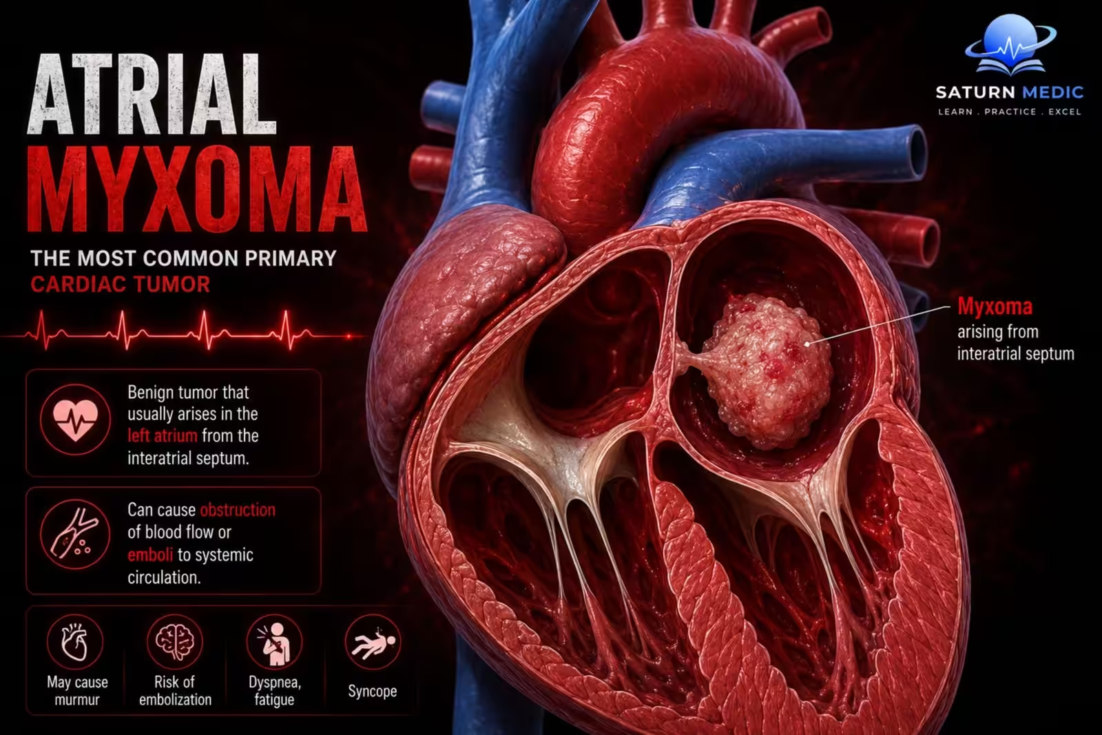
Cardiac myxoma is the most common primary tumor of the heart. It is usually benign and arises from primary connective tissue. Around 75% occur in the left atrium, usually attached to the interatrial septum. Right atrial myxomas are less common, while ventricular myxomas are rare. Symptoms mainly result from: Obstruction of intracardiac blood flow. Embolization of tumor fragments.
Etiology & Risk Factors
- Exact cause is unknown.
- Most cases are sporadic.
- About 10% are hereditary:
- Autosomal dominant inheritance
- Associated with Carney syndrome
Pathophysiology
-Gross pathology
- Usually pedunculated (attached by a stalk).
- Gelatinous in consistency.
-Microscopic pathology
- Scattered mesenchymal cells in a mucoid (gelatinous) matrix.
- Surrounded by glycosaminoglycans.
- Tumor cells produce VEGF.
-Mechanism of symptoms
- Obstruction of the mitral valve opening causes symptoms similar to mitral stenosis.
- Embolization may cause stroke or infarction in other organs.
- IL-6 production by the tumor leads to constitutional symptoms.
Clinical Presentation
-Constitutional symptoms
Weight loss
Fever
Pallor
Digital clubbing
-Symptoms due to obstruction
Dyspnea on exertion
Orthopnea
Paroxysmal nocturnal dyspnea
Palpitations
Dizziness
Recurrent syncope
-Physical examination
Low-pitched mid-diastolic rumbling murmur at the apex
Characteristic “tumor plop” (early diastolic sound)
Murmur and heart sounds may change with body position.
Mitral regurgitation may occur due to valve damage.
-Symptoms due to embolization
CNS: TIA, stroke, seizures
Abdomen: Visceral infarction or hemorrhage
Lungs: Pulmonary embolization
History Taking
Important points include:
- Progressive exertional dyspnea
- Episodes of syncope
- Palpitations
- Orthopnea or PND
- Constitutional symptoms (fever, weight loss)
- History suggestive of embolic events (stroke, TIA, limb or abdominal ischemia)
- Family history of Carney syndrome or hereditary cardiac myxoma
Investigations
1. Echocardiography — investigation of choice
- Transthoracic echocardiography (TTE) is usually the initial investigation.
- Demonstrates an intracardiac mass, typically arising from the left atrium.
- Assesses:
- Tumour size
- Site of attachment
- Mobility
- Obstruction of the mitral valve
- Effects on cardiac function
2. Transoesophageal echocardiography (TOE/TEE)
- Provides better definition of the tumour, its attachment and mobility, particularly when TTE is inadequate.
3. ECG
- May show non-specific abnormalities or atrial arrhythmias.
- Not diagnostic.
4. Chest X-ray
- May be normal.
- Can demonstrate cardiac enlargement or pulmonary venous congestion when significant obstruction is present.
5. Laboratory investigations
- May show inflammatory or constitutional abnormalities, including:
- Anaemia
- Raised inflammatory markers
- These findings are non-specific and support the clinical picture rather than establish the diagnosis.
6. Cardiac CT/MRI
- Can further characterise an intracardiac mass when echocardiography is inconclusive and help define its anatomical extent.
Diagnosis
-The diagnosis is primarily made by echocardiography.
-Typical diagnostic findings:
- Mobile atrial mass
- Usually located in the left atrium
- Often attached to the interatrial septum
- May prolapse through the mitral valve, producing functional obstruction.
–Differential diagnoses of an atrial mass include:
- Atrial thrombus
- Other cardiac tumours
- Vegetations
Management
1. Surgical excision — definitive treatment
- Prompt surgical removal is the treatment of choice.
- The tumour and its site of attachment/stalk are excised to reduce the risk of recurrence.
- Surgery is indicated because of the risks of:
- Embolisation
- Cardiac obstruction
- Heart failure
- Sudden cardiovascular deterioration
2. Pre-operative assessment
- Echocardiography defines the tumour and its haemodynamic effects.
- Cardiac imaging may be used to assess anatomy and plan surgery.
- Patients undergo standard assessment for cardiac surgery.
3. After surgery
- Histological examination confirms the diagnosis.
- Follow-up echocardiography is important because recurrence can occur, particularly in familial or syndromic cases.
Complications
- Systemic embolization
- Cerebral infarction (stroke)
- Pulmonary embolization
- Valvular damage causing mitral regurgitation
- Sudden death
- Tumor recurrence after incomplete excision or from another tumor focus
Prognosis
- Generally favorable after surgical resection.
- Recurrence can occur, especially after incomplete tumor removal.
Key Points / Clinical Pearls
- Most common primary benign cardiac tumor.
- About 75% arise in the left atrium, usually from the interatrial septum.
- Classic auscultatory finding: “tumor plop.”
- Symptoms are due to obstruction, embolization, and IL-6-mediated constitutional effects.
- Echocardiography is the investigation of choice.
- Immediate surgical excision is the definitive treatment because of the risk of embolization and sudden death.
- Vaidya Y, Sharma S. National Center for Biotechnology Information (NIH). Atrial Myxoma, StatPearls.
- Reynen K. Cardiac Myxomas. N Engl J Med. 1995;333:1610-1617. doi: 10.1056/NEJM199512143332407.
- Islam AKMM. Cardiac Myxomas: A Narrative Review. World J Cardiol. 2022;14:206-219. PMC9048271.
- MedlinePlus, National Library of Medicine (NIH). Atrial Myxoma: Medical Encyclopedia.
- Samanidis G, Khoury M, Balanika M, Perrea DN. Current Challenges in the Diagnosis and Treatment of Cardiac Myxoma. Kardiol Pol. 2020;78:269-277. doi: 10.33963/KP.15254.