

Clinical Subject Page
Atrioventricular Block (AV Block)
ICD-10
Specialty
Onset
Reviewed
-
OverviewOverview
-
Etiology & Risk FactorsEtiology & Risk Factors
-
PathophysiologyPathophysiology
-
Clinical PresentationClinical Presentation
-
History TakingHistory Taking
-
Degrees of AV BlockDegrees of AV Block
-
InvestigationsInvestigations
-
DiagnosisDiagnosis
-
ManagementManagement
-
ComplicationsComplications
-
PrognosisPrognosis
-
Key Points / Clinical PearlsKey Points / Clinical Pearls
Overview
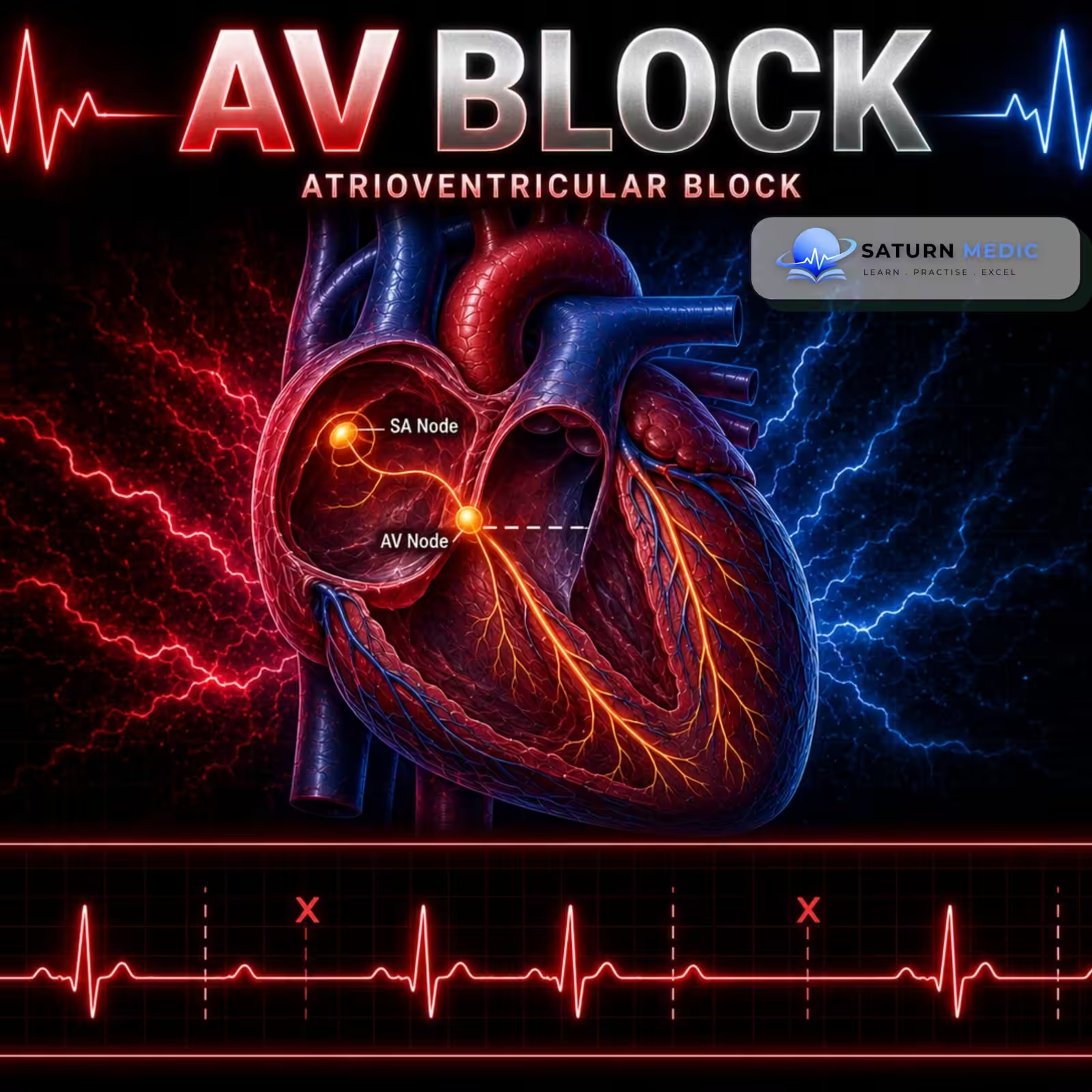
Atrioventricular (AV) block is a heart conduction disorder in which the electrical signals from the atria (upper chambers) to the ventricles (lower chambers) are delayed or completely blocked. This can cause a slow heart rate (bradycardia) and reduce blood flow to the body.
Etiology & Risk Factors
Cardiac Causes
Ischemic heart disease (especially acute myocardial infarction)
Congenital heart disease and congenital complete AV block
Post-cardiac procedures (e.g., valve surgery, catheter ablation, TAVI)
Myocarditis
Cardiomyopathies (e.g., amyloidosis, sarcoidosis)
Degenerative fibrosis of the conduction system (Lenègre-Lev syndrome)
Noncardiac Causes
Increased vagal tone (e.g., athletes, vomiting, suctioning, intubation)
Electrolyte disorders (especially hyperkalemia)
Acid-base disorders
Cardiotoxic drugs
Beta-blockers
Calcium channel blockers
Digoxin
Infections
Lyme carditis
Bacterial endocarditis
Acute rheumatic fever
Endocrine disorders
Thyroid disease
Adrenal disease
Neuromuscular disorders
Myotonic dystrophy
Risk Factors
Coronary artery disease or acute MI
Previous cardiac surgery or catheter ablation
Structural heart disease
Cardiomyopathy
Myocarditis
Electrolyte abnormalities (especially hyperkalemia)
Use of beta-blockers, calcium channel blockers, or digoxin
Lyme disease
High vagal tone (e.g., trained athletes)
Pathophysiology
- AV block occurs when the electrical impulse from the atria to the ventricles is interrupted or delayed anywhere in the AV conduction system, including:
- AV node
- Bundle of His
- Right and left bundle branches
- AV nodal block
- The impulse is delayed at the AV node.
- Ventricular conduction remains normal → narrow QRS complex.
- Infranodal block
- The block occurs at or below the Bundle of His.
- Ventricular depolarization is impaired → wide QRS complex.
- More distal (lower) blocks
- Result from more extensive damage to the conduction system.
- Have a higher risk of progressing to complete (third-degree) heart block.
Simple Flow
Damage to the AV conduction system → Delayed or blocked impulse from atria to ventricles → Slow ventricular rate (bradycardia) → ↓ Cardiac output → Dizziness, syncope, or heart failure
- AV block occurs when the electrical impulse from the atria to the ventricles is interrupted or delayed anywhere in the AV conduction system, including:
Clinical Presentation
Many Patients
Asymptomatic (especially first-degree and Mobitz type I AV block)
Common Symptoms (due to bradycardia)
Fatigue
Exercise intolerance
Shortness of breath (dyspnea)
Dizziness
Syncope (fainting)
Palpitations (especially with irregular rhythms such as Mobitz I)
Other Symptoms
Feeling of pulsations in the neck or chest (due to loss of AV synchrony)
Heart failure symptoms (fatigue, dyspnea, exercise intolerance)
Severe Presentation
Stokes-Adams attacks (sudden brief loss of consciousness due to ventricular asystole)
Cardiac arrest (especially in advanced AV block)
History Taking
- “Have you felt dizzy or lightheaded?”
- “Have you ever fainted or nearly fainted?”
- “Do you feel unusually tired?”
- “Do you get short of breath during activity?”
- “Do you notice your heart beating slowly or skipping beats?”
- “Do you have chest pain?”
- “Have your symptoms started suddenly or gradually?”
- “Have you had a recent heart attack or heart surgery?”
- “Are you taking medications such as beta-blockers, calcium channel blockers, or digoxin?”
- “Have you been diagnosed with Lyme disease or thyroid disease?”
Degrees of AV Block
Atrioventricular (AV) Block · Classification & Management
| Degree | ECG Findings | Typical Cause / Level of Block | Management |
|---|---|---|---|
| 1st Degree | PR > 200 ms fixed, all P waves conduct | Often AV node delay. Increased vagal tone, AV nodal disease, some drugs (beta-blockers, CCBs, digoxin). | No treatment usually required. Monitor; review/adjust contributing drugs if symptomatic. |
| 2nd Degree — Mobitz I | Progressive PR lengthening → dropped beat → resets | Usually AV node level. Increased vagal tone, inferior MI, drugs. | Usually benign and asymptomatic. Observe; treat reversible cause. Pacing rarely needed. |
| 2nd Degree — Mobitz II | Fixed PR, sudden non-conducted P waves, often fixed ratio | Infranodal (His-Purkinje/bundle branch) disease. Anterior MI, fibrosis, structural heart disease. | Higher risk of progression to complete block. Admit, cardiology referral, permanent pacemaker usually indicated even if asymptomatic. |
| High-Grade Block | ≥2 consecutive P waves not conducted, some conduction persists | Usually infranodal. Similar substrate to Mobitz II. | Treat as high risk — same urgency as Mobitz II/complete block. Pacing usually indicated. |
| 3rd Degree (Complete) | No P-QRS relationship, independent atrial & ventricular rates | AV node or infranodal. Can be congenital, degenerative (Lenegre/Lev), MI, post-cardiac surgery, infiltrative disease. | Medical emergency if unstable or symptomatic. Atropine/isoprenaline or transcutaneous pacing as bridge; permanent pacemaker definitive treatment. |
Investigations
1. 12-Lead ECG – First-line (Gold Standard)
Confirms the type and degree of AV block.
2. Continuous ECG Monitoring
Detects intermittent or high-grade AV block.
Used in symptomatic or hospitalized patients.
3. Laboratory Tests
Electrolytes (especially potassium)
Troponin (if myocardial infarction is suspected)
Renal function tests
Liver function tests
TSH
Blood gas (serum pH)
Lyme disease antibody testing (if suspected)
Drug levels (e.g., digoxin), when indicated
4. Cardiac Imaging
Transthoracic echocardiography (TTE) to evaluate structural heart disease.
Consider cardiac MRI, CT, or TEE if further evaluation is needed.
Diagnosis
Diagnostic Approach
12-lead ECG is the gold standard to diagnose and classify the degree of AV block.
Assess whether the patient is stable or unstable (signs of unstable bradycardia).
Identify the underlying cause (e.g., myocardial infarction, electrolyte abnormalities, medication toxicity).
ECG Findings
First-degree AV block: PR interval >200 ms.
Mobitz I (Wenckebach): Progressive PR prolongation → dropped QRS.
Mobitz II: Constant PR interval with intermittent dropped QRS complexes.
Third-degree AV block: AV dissociation (no relationship between P waves and QRS complexes).
Management
1. Initial Management
Obtain a 12-lead ECG immediately.
Assess for unstable bradycardia.
Apply transcutaneous pacing pads if needed.
2. Unstable Patients
IV atropine
Transcutaneous pacing
Transvenous pacing if required
IV chronotropic medication (e.g., epinephrine)
Stabilize first, then investigate the cause.
3. Stable Patients
Treat the underlying cause:
Correct electrolyte abnormalities.
Treat acute myocardial infarction.
Stop or adjust AV nodal-blocking drugs (e.g., beta-blockers, calcium channel blockers, digoxin).
Perform TTE if structural heart disease is suspected.
4. Low-Risk AV Block
(First-degree, Mobitz I, and some 2:1 AV blocks)
Usually observation and follow-up if asymptomatic.
Monitor with periodic ECGs.
Pacemaker only for selected symptomatic patients.
5. High-Risk AV Block
(Mobitz II, high-grade AV block, third-degree AV block)
Continuous cardiac monitoring
Temporary pacing if necessary.
Permanent pacemaker for most patients with irreversible AV block.
Complications
- Progression to complete (third-degree) heart block
- Symptomatic bradycardia
- Syncope (Stokes–Adams attacks)
- Heart failure (due to reduced cardiac output)
- Cardiac arrest
- Sudden cardiac death (especially with untreated high-grade AV block)
Prognosis
- First-degree AV block and Mobitz I generally have a good prognosis and often only require observation if asymptomatic.
- Mobitz II and third-degree AV block have a higher risk of progression and usually require permanent pacemaker implantation.
- Prognosis improves significantly when reversible causes are treated and high-grade AV block is managed with a permanent pacemaker.
Key Points / Clinical Pearls
- AV block = delayed or blocked electrical conduction from the atria to the ventricles.
- Three types: First-degree, Second-degree (Mobitz I & II), and Third-degree (complete heart block).
- Gold-standard diagnosis: 12-lead ECG.
- First-degree: PR interval >200 ms.
- Mobitz I: Progressive PR prolongation → dropped QRS.
- Mobitz II: Constant PR interval → dropped QRS (high risk).
- Third-degree: Complete AV dissociation.
- First-degree and Mobitz I: Usually observation if asymptomatic.
- Mobitz II and third-degree: Usually require a permanent pacemaker.
- Always identify and treat reversible causes (e.g., myocardial infarction, electrolyte abnormalities, medication toxicity).
- National Center for Biotechnology Information (NIH). Atrioventricular Block, StatPearls.
- Kusumoto FM, Schoenfeld MH, Barrett C, et al. 2018 ACC/AHA/HRS Guideline on the Evaluation and Management of Patients With Bradycardia and Cardiac Conduction Delay. Circulation. 2019;140:e382-e482. PMID: 30586772.
- MedlinePlus, National Library of Medicine (NIH). Heart Block: Medical Encyclopedia.
- National Center for Biotechnology Information (NIH). Second-Degree Atrioventricular Block, StatPearls.
- National Center for Biotechnology Information (NIH). Third-Degree Atrioventricular Block, StatPearls.