

Clinical Subject Page
Dilated Cardiomyopathy (DCMP)
Also called
ICD-10
Specialty
Onset
Reviewed
On This Page
-
OverviewOverview
-
Etiology & Risk FactorsEtiology & Risk Factors
-
PathophysiologyPathophysiology
-
Clinical PresentationClinical Presentation
-
History TakingHistory Taking
-
InvestigationsInvestigations
-
DiagnosisDiagnosis
-
ManagementManagement
-
ComplicationsComplications
-
PrognosisPrognosis
-
Key Points / Clinical PearlsKey Points / Clinical Pearls
Overview
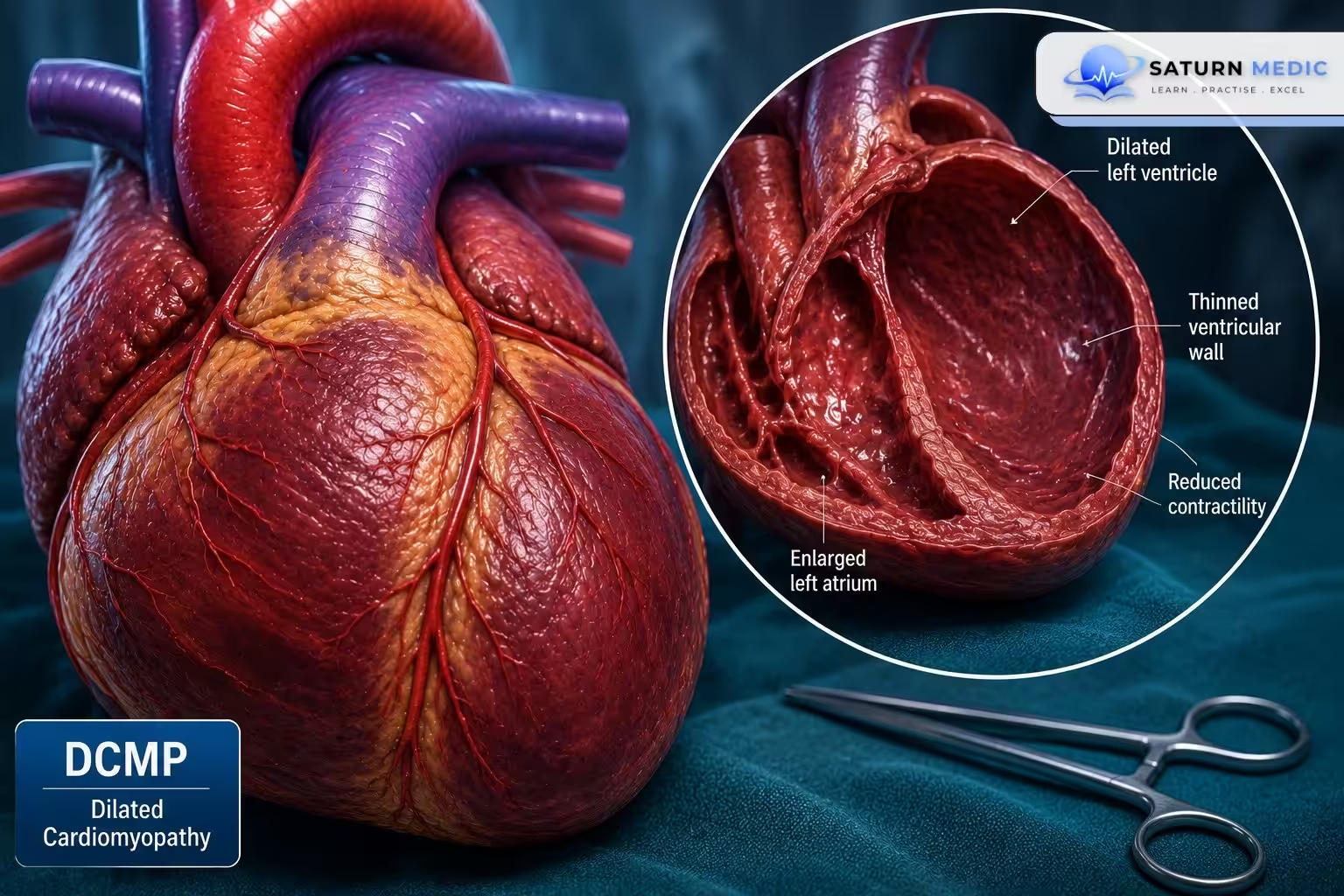
Dilated cardiomyopathy (DCM/DCMP) is a myocardial disease characterized by dilation of one or both ventricles with reduced systolic function (LVEF <40–50%), in the absence of abnormal loading conditions or coronary artery disease sufficient to explain the dysfunction.
Etiology & Risk Factors
Etiology of Dilated Cardiomyopathy :
- Idiopathic (most common)
- Genetic (familial DCM)
- Viral myocarditis (e.g., Coxsackie B, adenovirus)
- Alcohol abuse
- Chemotherapy (doxorubicin, trastuzumab)
- Peripartum cardiomyopathy
- Autoimmune diseases
- Endocrine disorders (thyroid disease, diabetes)
- Nutritional deficiencies (thiamine, selenium)
- Tachycardia-induced cardiomyopathy
Risk Factors for Dilated Cardiomyopathy :
- Family history of cardiomyopathy
- Hypertension
- Excessive alcohol intake
- Smoking
- Cocaine and illicit drug use
- Previous myocarditis
- Cancer chemotherapy
- Pregnancy (peripartum)
- Metabolic disorders
Pathophysiology
Myocardial injury/genetic defect
↓
Ventricular dilation
↓
Reduced contractility
↓
↓ Stroke volume & ↓ Ejection fraction
↓
Neurohormonal activation (RAAS & SNS)
↓
Fluid retention + Ventricular remodeling
↓
Heart failure (HFrEF)
↓
Mitral regurgitation + Arrhythmias + Thrombus formation
Clinical Presentation
Symptoms of Dilated Cardiomyopathy :
- Progressive exertional dyspnea
- Orthopnea
- Paroxysmal nocturnal dyspnea
- Fatigue
- Reduced exercise tolerance
- Peripheral edema
- Palpitations
- Syncope (advanced disease)
Signs of Dilated Cardiomyopathy :
- Tachycardia
- Elevated JVP
- S3 gallop
- Displaced, diffuse apex beat
- Functional mitral regurgitation murmur
- Bibasal crackles
- Hepatomegaly
- Peripheral edema
History Taking
Key Questions:
- When did the symptoms begin?
- Is dyspnea worsening?
- Orthopnea or PND?
- Chest pain?
- Palpitations?
- Syncope?
- Alcohol intake?
- Recreational drug use?
- Chemotherapy history?
- Recent viral illness?
- Family history of sudden cardiac death or cardiomyopathy?
Red Flags:
- Syncope
- Sustained ventricular arrhythmias
- Cardiogenic shock
- Sudden worsening heart failure
Investigations
Laboratory
- CBC
- Electrolytes
- Renal function
- Liver function
- BNP/NT-proBNP
- Troponin
- Thyroid function
- Iron studies
- Viral/autoimmune tests (selected patients)
ECG:
- Sinus tachycardia
- Left bundle branch block (LBBB)
- Atrial fibrillation
- Ventricular arrhythmias
- Nonspecific ST-T changes
Imaging:
- Chest X-ray
- Cardiomegaly
- Pulmonary congestion
- Echocardiography (gold standard)
- Dilated LV
- Reduced LVEF
- Global hypokinesia
- Functional MR
- Cardiac MRI
- Myocardial fibrosis
- Myocarditis evaluation
Additional Tests:
- Coronary angiography/CT coronary angiography
- Genetic testing (familial disease)
- Endomyocardial biopsy (selected cases)
Diagnosis
Diagnosis is based on:
- Clinical features of heart failure
- Echocardiographic evidence of ventricular dilation with reduced systolic function
- Exclusion of significant coronary artery disease or abnormal loading conditions
- Identification of an underlying cause when possible
Dilated Cardiomyopathy (DCM) · Diagnostic Criteria
Management
Lifestyle
- Salt restriction
- Fluid restriction (selected patients)
- Alcohol cessation
- Smoking cessation
- Regular exercise as tolerated
- Vaccinations
- Family screening if inherited
Medications (Guideline-Directed Medical Therapy)
- ARNI (preferred) or ACE inhibitor/ARB
- Evidence-based β-blocker
- Mineralocorticoid receptor antagonist (MRA)
- SGLT2 inhibitor
- Loop diuretics for congestion
- Anticoagulation if atrial fibrillation or LV thrombus
Devices
- ICD for prevention of sudden cardiac death
- CRT for selected patients with LBBB and reduced EF
Advanced Therapy
- LVAD
- Heart transplantation
Complications
- Heart failure progression
- Atrial fibrillation
- Ventricular tachycardia/fibrillation
- Sudden cardiac death
- Functional mitral regurgitation
- Left ventricular thrombus
- Stroke/systemic embolism
- Cardiogenic shock
Prognosis
- Variable depending on etiology and response to therapy
- Many patients improve with modern GDMT
- Poor prognosis with severe LV dysfunction or recurrent ventricular arrhythmias
- Advanced disease may require transplantation
Key Points / Clinical Pearls
- Dilated Cardiomyopathy is the most common cause of HFrEF.
- Echocardiography is the first-line imaging modality.
- Exclude ischemic heart disease before diagnosing idiopathic DCM.
- Early guideline-directed medical therapy improves survival.
- ICD reduces the risk of sudden cardiac death in eligible patients.
- Consider genetic testing and family screening in familial Dilated Cardiomyopathy.
- Mahmaljy H, Yelamanchili VS, Singhal M. National Center for Biotechnology Information (NIH). Dilated Cardiomyopathy, StatPearls.
- Arbelo E, Protonotarios A, Gimeno JR, et al. 2023 ESC Guidelines for the Management of Cardiomyopathies. Eur Heart J. 2023;44:3503-3626. doi: 10.1093/eurheartj/ehad194.
- MedlinePlus, National Library of Medicine (NIH). Dilated Cardiomyopathy: Medical Encyclopedia.
- Diagnosis and Management of Dilated Cardiomyopathy: A Systematic Review of Clinical Practice Guidelines and Recommendations. Eur Heart J. PMID: 39674807.
- Heidenreich PA, Bozkurt B, Aguilar D, et al. 2022 AHA/ACC/HFSA Guideline for the Management of Heart Failure. Circulation. 2022;145:e895-e1032. PMID: 35363499.